

Clinical Subject Page
Mitral Stenosis
Also called
ICD-10
Specialty
Onset
Reviewed
-
OverviewOverview
-
Etiology & Risk FactorsEtiology & Risk Factors
-
PathophysiologyPathophysiology
-
Clinical PresentationClinical Presentation
-
History TakingHistory Taking
-
Mitral Stenosis Vs Mitral RegurgitationMitral Stenosis Vs Mitral Regurgitation
-
InvestigationsInvestigations
-
DiagnosisDiagnosis
-
ManagementManagement
-
ComplicationsComplications
-
PrognosisPrognosis
-
Key Points / Clinical PearlsKey Points / Clinical Pearls
Overview
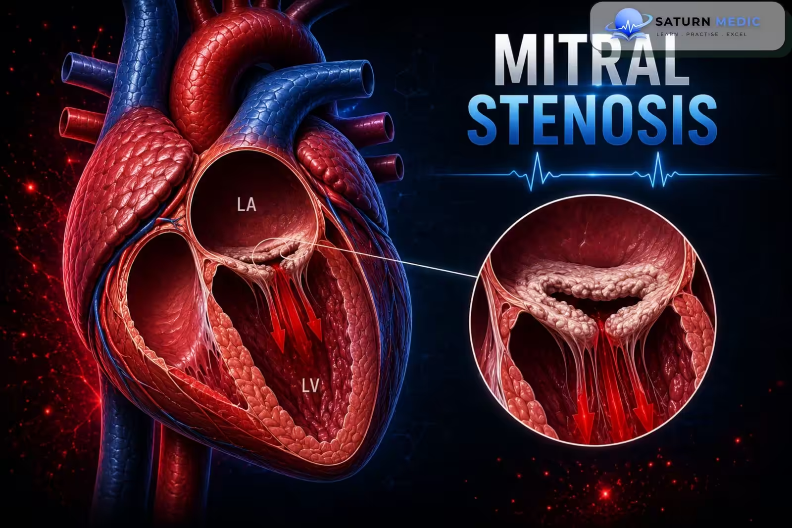
Mitral stenosis (MS) is a narrowing of the mitral valve, which reduces blood flow from the left atrium to the left ventricle. This causes increased pressure in the left atrium, leading to pulmonary congestion, pulmonary hypertension, and eventually heart failure if untreated.
Etiology & Risk Factors
Most Common Cause
Rheumatic fever (most common cause worldwide)
Other Causes
Calcification of the mitral valve annulus
Autoimmune diseases
Systemic lupus erythematosus (SLE)
Rheumatoid arthritis (RA)
Congenital mitral stenosis
Conditions that may mimic MS:
Bacterial endocarditis with large mitral valve vegetation
Left atrial myxoma
Degenerative aortic stenosis (may reduce the mitral annular area)
Risk Factors
History of rheumatic fever (most important risk factor)
Increasing age (risk of mitral annular calcification)
Autoimmune diseases (e.g., SLE, RA)
Congenital heart disease
Pathophysiology
- Mitral valve stenosis causes obstruction of blood flow from the left atrium (LA) to the left ventricle (LV).
- This results in reduced LV filling during diastole → decreased stroke volume → decreased cardiac output (forward heart failure).
- Blood backs up into the left atrium, causing increased left atrial pressure.
- The increased pressure is transmitted to the pulmonary veins and lungs, leading to:
- Pulmonary venous congestion
- Cardiogenic pulmonary edema
- Pulmonary hypertension
- Long-standing pulmonary hypertension can cause right ventricular hypertrophy and right-sided (backward) heart failure.
Simple Flow
Mitral valve narrowing → Obstruction of LA → LV blood flow → ↑ Left atrial pressure → Pulmonary congestion → Pulmonary hypertension → Right ventricular hypertrophy → Right-sided heart failure
Clinical Presentation
Common Symptoms
Dyspnea (shortness of breath) – most common symptom
Fatigue
Palpitations (often due to atrial fibrillation)
Orthopnea
Paroxysmal nocturnal dyspnea (PND)
Hemoptysis
Hoarseness (Ortner syndrome)
Dysphagia
Symptoms of embolic disease (e.g., stroke)
Late Features
Symptoms of right-sided heart failure
Leg swelling (edema)
Increasing breathlessness
Physical Examination
Irregular pulse (if atrial fibrillation is present)
Diastolic murmur heard best at the apex
Loud first heart sound (S1)
Opening snap after S2
Signs of right heart failure (e.g., peripheral edema)
History Taking
- “Do you get short of breath when walking or climbing stairs?”
- “Do you become breathless when lying flat?”
- “Do you wake up at night feeling short of breath?”
- “Do you feel your heart racing or skipping beats?”
- “Do you feel tired more easily than before?”
- “Have you coughed up blood?”
- “Have you noticed swelling in your legs or ankles?”
- “Have you ever had rheumatic fever or recurrent sore throats?”
- “Have you ever had a stroke or sudden weakness?”
Mitral Stenosis Vs Mitral Regurgitation
Cardiology · Valvular Heart Disease — Mitral Stenosis vs Mitral Regurgitation
Normal area: 4–6 cm² | Severe: <1 cm²
Most common cause: Rheumatic fever
Volume overload of both LA and LV
Most common cause: MVP / rheumatic / ischaemic
Timing Diastolic — after S2, continues toward S1
Radiation Does not radiate
Character Low-pitched, rumbling
Special Loud S1 | Opening snap after S2
Positional Louder in left lateral decubitus
S1┈┈┈S2┈OS┈╾╾╾rumble╿╿╿┈S1
└ Shorter S2–OS interval = more severe stenosis (higher LA pressure)
Timing Throughout systole — S1 to S2
Radiation Axilla / left infrascapular
Character Blowing, high-pitched
Special Soft/absent S1 | S3 gallop (volume overload)
Positional Louder with squatting & expiration
S1┈╾╾╾╾╾╾╾╾╾S2┈┈┈┈┈┈S1
└ Uniform throughout systole — does not change with S1 or S2
P in V1 Biphasic — prominent negative terminal deflection
QRS Usually normal (LV not overloaded)
Late finding RVH pattern (RAD, dominant R in V1) if pulmonary HTN
Rhythm Atrial fibrillation common (LA dilation)
┈/╾╿┈P-mitrale┈┈┈┈narrow QRS┈┈┈┈
└ P mitrale = LA enlargement signature — notched P >2.5 mm wide in II
QRS LVH pattern — tall R in V5/V6, deep S in V1/V2
Axis LAD (LV volume overload)
Rhythm AF common (LA dilation, same as MS)
Key diff LVH present in MR — absent in MS
┈/╾╿┈P-mitrale┈┈tall QRS (LVH)┈┈
└ LVH on ECG = volume overload — key distinguisher from pure MS
| Feature | Mitral Stenosis | Mitral Regurgitation |
|---|---|---|
| Murmur timing | Mid-diastolic rumble | Pansystolic blowing |
| Best heard | Apex, left lateral decubitus | Apex, radiates to axilla |
| S1 | Loud (early) → soft (late/calcified) | Soft / obscured |
| Extra sounds | Opening snap (OS) after S2 | S3 gallop (volume overload) |
| ECG P wave | P mitrale (broad, notched) | P mitrale (broad, notched) |
| ECG QRS | Normal (RVH if pulm HTN) | LVH pattern |
| CXR heart size | LA enlarged, LV normal | LA + LV both enlarged |
| Pulmonary oedema | Chronic (gradual venous HTN) | Acute (sudden decompensation) |
| LV function | Preserved (underfilled) | Dilated → EF may fall |
| AF risk | Very high (thrombus → stroke) | High (LA dilation) |
| Main cause | Rheumatic fever | MVP, rheumatic, ischaemic, IE |
| Intervention | PTMC (balloon) or MVR | Repair preferred over replacement |
| Surgery trigger | MVA <1.5 cm² + symptoms | EF ≤60% or ESD ≥40 mm |
Investigations
1. Transthoracic Echocardiography (TTE) – Gold Standard
Most important test for diagnosis and treatment planning.
Shows:
Reduced mitral valve area (MVA) (≤ 1.5 cm² = severe MS)
Thickened, calcified mitral leaflets
Commissural fusion
Increased diastolic pressure gradient
Left atrial enlargement
Pulmonary hypertension
Right ventricular dilation
2. ECG
May be normal early.
Can show:
Left atrial enlargement (P mitrale)
Atrial fibrillation
Right ventricular hypertrophy (late disease)
3. Chest X-ray
Left atrial enlargement
Pulmonary congestion
Right ventricular enlargement
4. Laboratory Tests
BNP/NT-proBNP
CBC
BMP
Liver function tests
CRP (if rheumatic heart disease is suspected)
5. Additional Tests (when indicated)
Transesophageal echocardiography (TEE) before intervention or if TTE is inconclusive
Exercise stress test if symptoms do not match TTE findings
Cardiac catheterization if disease severity is unclear or before surgery
Diagnosis
Diagnostic Approach
Suspect MS based on symptoms (e.g., dyspnea, fatigue, palpitations) and physical examination.
Confirm the diagnosis with transthoracic echocardiography (TTE).
Gold-Standard Test:
Transthoracic Echocardiography (TTE)
Best initial and most important test for diagnosing MS.
Typical findings:
Reduced mitral valve area (MVA)
MVA ≤ 1.5 cm² = severe mitral stenosis
Thickened, calcified mitral valve leaflets with commissural fusion
Increased mean diastolic pressure gradient
Left atrial enlargement
Pulmonary hypertension
Right ventricular dilation
Supportive Tests
ECG
Left atrial enlargement (P mitrale)
Atrial fibrillation
Right ventricular hypertrophy (late finding)
Chest X-ray
Left atrial enlargement
Pulmonary congestion
Right ventricular enlargement
TEE
If TTE is inconclusive
Before intervention to detect left atrial thrombus or significant mitral regurgitation
Management
1. Medical Management
Asymptomatic patients: Regular follow-up with TTE.
Treat heart failure symptoms and associated conditions.
Manage atrial fibrillation and other complications if present.
2. Interventional Management
Indications:
Symptomatic severe MS (MVA ≤ 1.5 cm²)
Asymptomatic severe MS with:
Pulmonary artery systolic pressure >50 mmHg, or
New-onset atrial fibrillation
3. Procedures
Percutaneous Mitral Balloon Commissurotomy (PMBC)
Preferred treatment for most patients with severe MS.
Balloon is used to open the narrowed mitral valve.
Surgery
Open commissurotomy or mitral valve replacement
Indicated if:
Anatomy is unsuitable for PMBC
Left atrial thrombus is present
Significant associated valve disease (e.g., severe mitral regurgitation)
Complications
- Atrial fibrillation (AF) → increases the risk of thromboembolic events (e.g., stroke)
- Pulmonary edema
- Pulmonary hypertension
- Congestive heart failure (CHF)
- Enlarged left atrium causing:
- Esophageal compression → dysphagia
- Recurrent laryngeal nerve palsy → hoarseness (Ortner syndrome)
Prognosis
- Mitral stenosis is slowly progressive, and many patients remain asymptomatic for years.
- Prognosis worsens as the valve becomes more narrowed and complications such as atrial fibrillation, pulmonary hypertension, and heart failure develop.
- Early diagnosis, regular follow-up, and timely intervention (e.g., balloon commissurotomy or surgery) improve outcomes
Key Points / Clinical Pearls
- Most common cause: Rheumatic fever
- Gold-standard test: Transthoracic echocardiography (TTE)
- Classic murmur: Opening snap followed by a diastolic murmur at the apex
- Most common symptom: Dyspnea
- Severe MS: Mitral valve area ≤ 1.5 cm²
- Preferred treatment for severe symptomatic MS: Percutaneous mitral balloon commissurotomy (PMBC)
- Common complications: Atrial fibrillation, stroke, pulmonary hypertension, and heart failure
- National Center for Biotechnology Information (NIH). Mitral Stenosis, StatPearls.
- Otto CM, Nishimura RA, Bonow RO, et al. 2020 ACC/AHA Guideline for the Management of Patients With Valvular Heart Disease. Circulation. 2021;143:e72-e227. PMID: 33972115.
- Haleem SM, Ahmed I, Kanmanthareddy A. National Center for Biotechnology Information (NIH). Catheter-Based Management of Mitral Stenosis, StatPearls.
- National Center for Biotechnology Information (NIH). Rheumatic Heart Disease, StatPearls.
- MedlinePlus, National Library of Medicine (NIH). Mitral Stenosis: Medical Encyclopedia.