

Clinical Subject Page
Pericarditis
Also called
ICD-10
Specialty
Onset
Reviewed
-
OverviewOverview
-
Etiology & Risk FactorsEtiology & Risk Factors
-
PathophysiologyPathophysiology
-
Clinical PresentationClinical Presentation
-
History TakingHistory Taking
-
InvestigationsInvestigations
-
DiagnosisDiagnosis
-
ManagementManagement
-
ComplicationsComplications
-
PrognosisPrognosis
-
Key Points / Clinical PearlsKey Points / Clinical Pearls
Overview
Pericarditis is inflammation of the pericardium (the fibrous sac surrounding the heart). It may be: Acute: lasts from days to weeks Chronic: persists for more than 3 months Acute pericarditis is most commonly caused by a viral infection, although many other conditions can trigger inflammation of the pericardium.
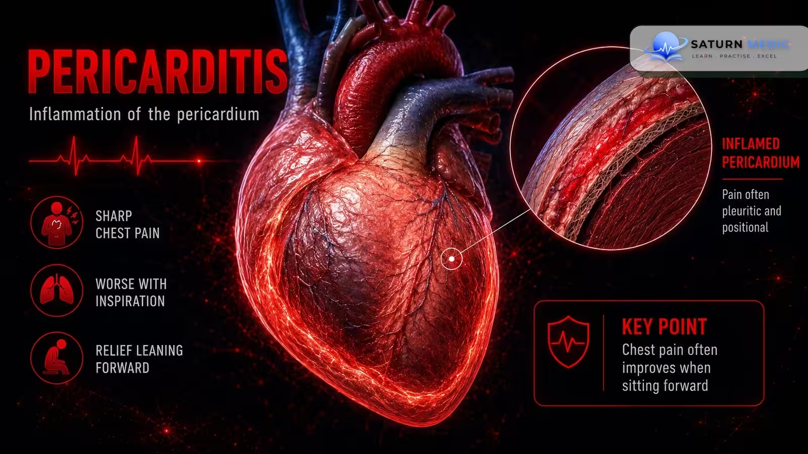
Typical features include: Low-grade fever Pleuritic chest pain (worse with deep inspiration) Pericardial friction rub on auscultation
Etiology & Risk Factors
-Etiology
Common causes include:
1. Idiopathic
- No identifiable cause (many are presumed viral)
2. Infectious
- Viral (most common): e.g., Coxsackie B virus
- Bacterial (e.g., Staphylococcus, Streptococcus, Mycobacterium tuberculosis)
- Fungal infections
- Toxoplasmosis
3. Cardiac causes
- Myocardial infarction
- Early post-infarction fibrinous pericarditis (within 1–3 days)
- Dressler syndrome (weeks to months after MI)
4. Postoperative/Traumatic
- Postpericardiotomy syndrome
- Blunt or penetrating chest trauma
5. Metabolic
- Uremia due to acute or chronic renal failure
6. Radiation-induced
7. Malignancy
- e.g., Hodgkin lymphoma
8. Autoimmune diseases
- Rheumatoid arthritis
- Systemic lupus erythematosus
- Scleroderma
9. Rare cause
- Following mRNA COVID-19 vaccination (rare; risk highest in males aged 12–24 years, and the risk is higher after SARS-CoV-2 infection than after vaccination)
-Risk Factors
Based on the reported causes, factors associated with an increased risk include:
- Viral or other infectious illnesses
- Recent myocardial infarction
- Recent cardiac surgery or chest trauma
- Acute or chronic kidney failure (uremia)
- Previous chest radiation therapy
- Autoimmune connective tissue diseases
- Malignancy
- Rarely, recent mRNA COVID-19 vaccination (especially in young males)
Pathophysiology
Pericarditis develops when the pericardium becomes inflamed due to infection or another inflammatory trigger.
The inflammatory process can:
- Cause inflammation of the pericardial layers.
- Produce chest pain because the inflamed pericardial surfaces rub against each other.
- Lead to accumulation of fluid in the pericardial space (pericardial effusion) in some patients.
If significant pericardial fluid accumulates, it may progress to cardiac tamponade, in which:
- Pericardial fluid increases pressure within the pericardial sac.
- The heart, especially the thin-walled right ventricle, is compressed.
- Ventricular filling during diastole decreases.
- Stroke volume and cardiac output decrease.
- Venous congestion develops, potentially causing life-threatening hemodynamic compromise.
Clinical Presentation
Pericarditis · Clinical Presentation
History Taking
1. Chest Pain
- Where is the pain?
- Is it sharp or stabbing?
- Does it worsen with deep breathing or coughing?
- Does it improve when sitting up or leaning forward?
- Does it radiate to the neck or left shoulder?
2. Associated Symptoms
- Do you have fever?
- Are you short of breath?
- Do you have a dry cough?
- Have you noticed palpitations or fainting?
3. Possible Causes
- Have you had a recent viral illness or fever?
- Have you had a recent heart attack, heart surgery, or chest injury?
- Do you have kidney disease, autoimmune disease, or cancer?
4. Red Flags
- Is your fever above 38°C?
- Is your shortness of breath getting worse?
- Have you ever fainted?
Investigations
1. Electrocardiogram (ECG) – First-line
Typical findings:
Diffuse ST-segment elevation
PR-segment depression
Later: T-wave inversion
2. Echocardiography (TTE) – First-line
Detects pericardial effusion
Assesses for cardiac tamponade
May be normal in uncomplicated pericarditis
3. Blood Tests
CBC: leukocytosis
CRP and ESR: elevated inflammatory markers
Troponin I/T: may be mildly elevated (higher levels suggest associated myocarditis)
Creatine kinase (CK): may be elevated
4. Chest X-ray
Usually normal
May show an enlarged cardiac silhouette if there is a large pericardial effusion
5. CT Scan or Cardiac MRI (if diagnosis is uncertain)
Detects pericardial inflammation
Shows pericardial thickening or effusion
6. Pericardiocentesis (if indicated)
Indications:
Cardiac tamponade
Large pericardial effusion
Suspected bacterial or malignant pericarditis
Pericardial fluid analysis:
Gram stain and culture
Acid-fast bacilli (TB) testing
PCR
Cytology
Additional Tests (Based on Suspected Cause)
Kidney function: BUN, creatinine, electrolytes (uremia)
Blood cultures (suspected bacterial infection)
ANA and rheumatoid factor (autoimmune disease)
TB testing and HIV test (suspected tuberculous pericarditis)
Diagnosis
| Feature | Acute Pericarditis | Chronic Pericarditis (Constrictive) |
|---|---|---|
| Diagnostic basis | Diagnosis requires ≥ 2 of 4 criteria | Diagnosis is based primarily on characteristic imaging findings |
| Clinical findings | Characteristic pleuritic chest pain and/or pericardial friction rub | Symptoms and signs of constrictive physiology (assessed with imaging) |
| ECG | Diffuse ST-segment elevation, PR-segment depression | May show low QRS voltage, generalized flat/inverted T waves; atrial fibrillation may occur |
| Echocardiography (TTE) | Detects new/worsening pericardial effusion; may be normal | Increased pericardial thickness, abnormal ventricular filling, respiratory variation in filling |
| CT / Cardiac MRI | Used if diagnosis is uncertain; shows pericardial inflammation, thickening, or effusion | Shows pericardial thickening (>2 mm) and calcification; confirms constrictive disease |
| Laboratory tests | CBC, CRP, ESR, Troponin I/T | Similar tests as acute; BNP may help differentiate from restrictive cardiomyopathy, LFTs may be abnormal |
| Additional tests | Pericardiocentesis if tamponade, large effusion, or suspected bacterial/malignant cause | Cardiac catheterization if noninvasive tests are inconclusive |
Management
| Management | Acute Pericarditis | Chronic Pericarditis (Constrictive) |
|---|---|---|
| First-line treatment | NSAIDs (aspirin, ibuprofen, or indomethacin) | Consider anti-inflammatory therapy if transient constrictive pericarditis |
| Adjunct therapy | Colchicine with NSAIDs or as monotherapy | Treat heart failure (e.g., diuretics, sodium restriction) |
| Corticosteroids | Prednisone only in severe cases or when caused by uremia, connective tissue disease, or autoreactivity | May be considered if inflammation is present |
| Treat underlying cause | Antibiotics (bacterial), TB therapy, immunosuppressants (autoimmune), dialysis (uremia) | Treat the underlying cause whenever identified |
| Activity | Restrict physical activity until symptoms and inflammatory markers resolve | Activity according to heart failure symptoms |
| Surgical treatment | Pericardiocentesis for cardiac tamponade or large pericardial effusion | Pericardiectomy for persistent symptomatic constrictive or effusive-constrictive pericarditis |
| Hospital admission | Required for poor prognosis (e.g., fever >38°C, large effusion, tamponade, elevated troponin, immunosuppression, anticoagulant therapy, recurrent disease, or failure to respond to NSAIDs) | Consider admission if heart failure symptoms are present or further evaluation is required |
Complications
| Complication | Description |
|---|---|
| Cardiac tamponade | Accumulation of pericardial fluid causing compression of the heart, impaired ventricular filling, and reduced cardiac output. This is a medical emergency. |
| Constrictive pericarditis | Chronic scarring and thickening of the pericardium that restricts ventricular filling, leading to symptoms of right-sided heart failure. |
Prognosis
| Condition | Prognosis |
|---|---|
| Acute Pericarditis | Usually self-limited and resolves within days to weeks with appropriate anti-inflammatory treatment (NSAIDs ± colchicine). Most patients without poor prognostic features can be managed as outpatients. |
| Chronic Pericarditis | Defined as inflammation lasting > 3 months. Chronic disease is uncommon (about 2% of acute cases) and may progress to constrictive or effusive-constrictive pericarditis, which can require pericardiectomy. |
Poor Prognostic Features (Acute Pericarditis)
- Fever > 38°C
- Subacute onset
- Large pericardial effusion
- Elevated troponin
- Immunosuppression
- Anticoagulant therapy
- Recurrent pericarditis
- Signs of hemodynamic compromise (e.g., hypotension, jugular venous distention)
- No response to NSAIDs after 1 week
Key Points / Clinical Pearls
- Acute pericarditis is most commonly caused by a viral infection.
- Classic chest pain is sharp, pleuritic, worsens with inspiration, and improves when sitting up and leaning forward.
- Pericardial friction rub is the characteristic physical examination finding.
- Diagnosis of acute pericarditis requires ≥ 2 of the following:
- Characteristic chest pain
- Pericardial friction rub
- Typical ECG changes (diffuse ST-segment elevation)
- New or worsening pericardial effusion
- ECG typically shows diffuse ST-segment elevation and PR-segment depression.
- Echocardiography (TTE) is the first-line imaging study to detect pericardial effusion and assess for cardiac tamponade.
- NSAIDs plus colchicine are the mainstay of treatment for most patients with acute pericarditis.
- Cardiac tamponade is the most serious acute complication and requires urgent pericardiocentesis.
- Constrictive pericarditis is the major chronic complication and may require pericardiectomy.
- Poor prognostic features include fever > 38°C, large pericardial effusion, elevated troponin, recurrent disease, immunosuppression, anticoagulant therapy, and hemodynamic compromise.
- Dababneh E, Siddique MS. National Center for Biotechnology Information (NIH). Pericarditis, StatPearls.
- Schulz-Menger J, Collini V, Gröschel J, et al. 2025 ESC Guidelines for the Management of Myocarditis and Pericarditis. Eur Heart J. 2025;46:3952-4041. doi: 10.1093/eurheartj/ehaf192.
- Imazio M, Brucato A, Cemin R, et al; ICAP Investigators. A Randomized Trial of Colchicine for Acute Pericarditis. N Engl J Med. 2013;369:1522-1528. PMID: 23992557.
- MedlinePlus, National Library of Medicine (NIH). Pericarditis: Medical Encyclopedia.
- Klein AL, Imazio M, Cremer P, et al. Phase 3 Trial of Interleukin-1 Trap Rilonacept in Recurrent Pericarditis. N Engl J Med. 2021;384:31-41. PMID: 33200890.