

Clinical Subject Page
Pulmonary Hypertension
ICD-10
Specialty
Onset
Reviewed
-
OverviewOverview
-
Etiology & Risk FactorsEtiology & Risk Factors
-
PathophysiologyPathophysiology
-
Clinical PresentationClinical Presentation
-
History TakingHistory Taking
-
ClassificationClassification
-
InvestigationsInvestigations
-
DiagnosisDiagnosis
-
ManagementManagement
-
ComplicationsComplications
-
PrognosisPrognosis
-
Key Points / Clinical PearlsKey Points / Clinical Pearls
Overview
Pulmonary hypertension (PH) is a hemodynamic disorder characterized by elevated pressure in the pulmonary circulation, leading to increased right ventricular afterload, right ventricular dysfunction, and, if untreated, right heart failure.
Etiology & Risk Factors
WHO Group 1 – Pulmonary Arterial Hypertension (PAH)
- Idiopathic
- Heritable (BMPR2 mutation)
- Drug/toxin-induced
- Connective tissue diseases
- Congenital heart disease
- Portal hypertension
- HIV infection
WHO Group 2 – Due to Left Heart Disease
- Heart failure (HFrEF/HFpEF)
- Mitral or aortic valve disease
- Cardiomyopathy
WHO Group 3 – Due to Lung Disease/Hypoxia
- COPD
- Interstitial lung disease
- Obstructive sleep apnea
- Chronic hypoxemia
WHO Group 4 – Chronic Thromboembolic PH (CTEPH)
- Chronic pulmonary embolism
WHO Group 5 – Multifactorial
- Sarcoidosis
- Hematologic disorders
- Chronic kidney disease
- Other systemic disorders
Risk Factors
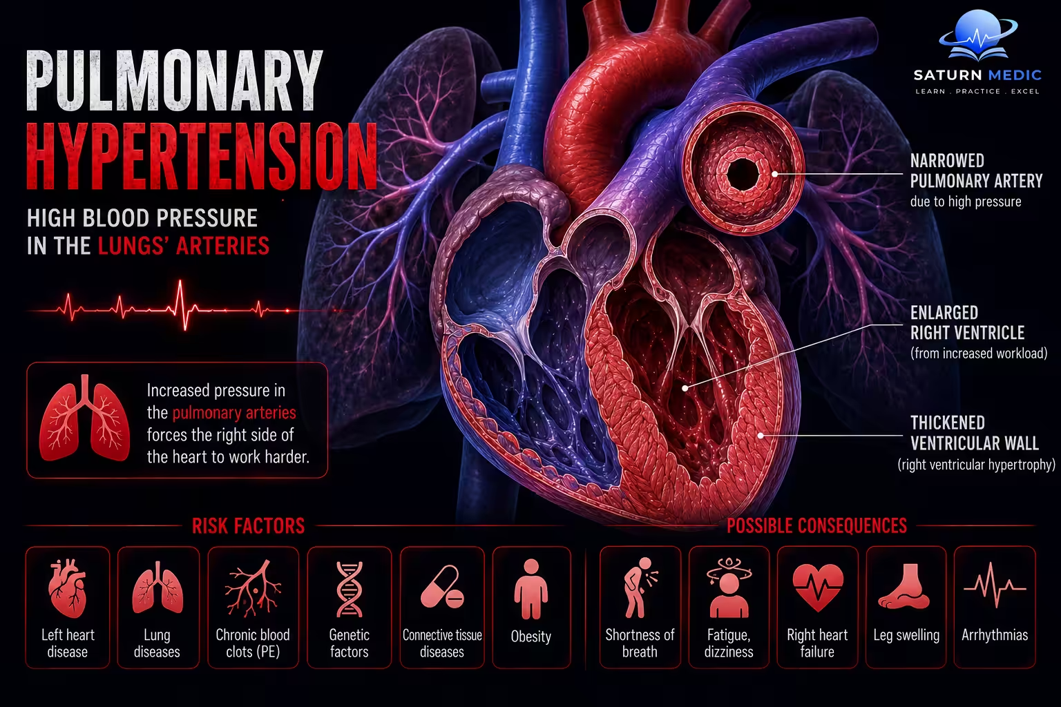
- Connective tissue disease
- Congenital heart disease
- Chronic lung disease
- Pulmonary embolism
- Family history
- HIV infection
- Portal hypertension
- Appetite suppressants or methamphetamine use
- Obesity and sleep apnea
Pathophysiology
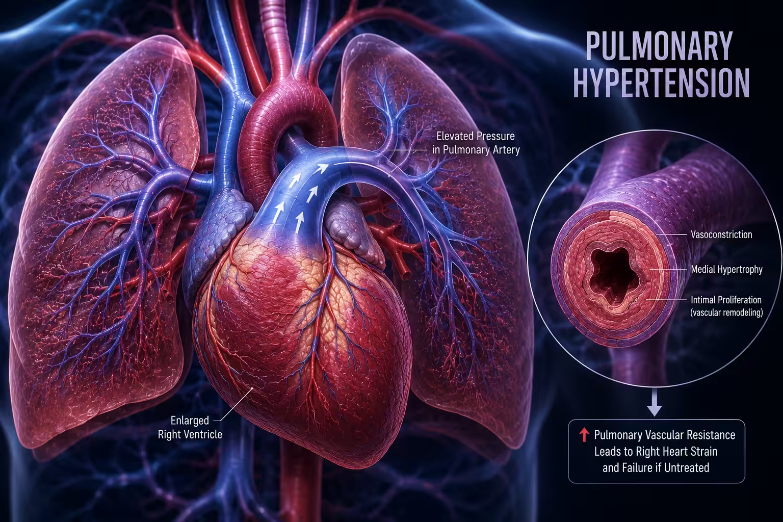
Pulmonary hypertension develops when there is an increase in pulmonary vascular resistance, pulmonary venous pressure, or pulmonary blood flow, leading to elevated pressure in the pulmonary circulation.
Mechanisms
- Increased pulmonary vascular resistance
- Occlusive vasculopathy
- Chronic hypoxic pulmonary vasoconstriction
- Endothelial dysfunction:
- ↑ Endothelin
- ↓ Nitric oxide (NO)
- ↓ Prostacyclin
- Inflammation causing vascular remodeling and fibrosis
- Increased pulmonary venous pressure
- Due to left-sided heart disease (e.g., mitral valve disease)
- Increased pulmonary blood flow
- Left-to-right shunts (e.g., ASD, VSD, PDA)
- Portopulmonary hypertension
- Sickle cell disease
Simple Flow
↑ Pulmonary vascular resistance / ↑ Pulmonary venous pressure / ↑ Pulmonary blood flow → ↑ Pulmonary artery pressure → ↑ Right ventricular afterload → Right ventricular hypertrophy and dilation → Right-sided heart failure (cor pulmonale) → Arrhythmias
Clinical Presentation
Symptoms
Progressive exertional dyspnea
Fatigue
Chest pain
Palpitations
Dizziness
Syncope (advanced disease)
Peripheral edema
Abdominal distension
Signs
Loud P2
Right ventricular heave
Elevated JVP
Tricuspid regurgitation murmur
Peripheral edema
Hepatomegaly
Ascites
Cyanosis (advanced disease)
History Taking
- When did the shortness of breath begin?
- Does it occur with exertion?
- Chest pain or syncope?
- Lower limb swelling?
- Previous pulmonary embolism?
- History of COPD or ILD?
- Congenital heart disease?
- Autoimmune disease?
- Family history of PH?
- Drug or appetite suppressant use?
Red Flags
- Syncope
- Resting dyspnea
- Chest pain
- Rapid symptom progression
- Signs of right heart failure
Classification
Pulmonary Hypertension · WHO Classification
| WHO Group | Haemodynamic Pattern | Key Examples | Treatment Approach |
|---|---|---|---|
| Group 1 — PAH | Pre-capillary mPAP >20, PVR >2 WU, PAWP ≤15 mmHg | Idiopathic PAH, scleroderma-associated, congenital heart disease (Eisenmenger), drug-induced | PAH-specific therapy — ERAs, PDE5 inhibitors, prostacyclin analogues, soluble guanylate cyclase stimulators |
| Group 2 — Left Heart Disease | Post-capillary mPAP >20, PAWP >15 mmHg | HFpEF/HFrEF, mitral/aortic valve disease | Treat underlying cardiac disease (diuretics, GDMT); PAH-specific drugs generally avoided/harmful |
| Group 3 — Lung Disease/Hypoxia | Pre-capillary mPAP >20, PAWP ≤15 mmHg | COPD, idiopathic pulmonary fibrosis, combined pulmonary fibrosis-emphysema, OSA | Optimise underlying lung disease, long-term oxygen therapy; PAH drugs not routinely recommended |
| Group 4 — CTEPH | Pre-capillary mPAP >20, PAWP ≤15 mmHg | Chronic thromboembolic PH after incomplete clot resolution; rare pulmonary artery tumour/sarcoma | Pulmonary endarterectomy (potentially curative); balloon pulmonary angioplasty or riociguat if inoperable |
| Group 5 — Unclear/Multifactorial | Variable — mixed pre-/post-capillary mechanisms | Sarcoidosis, chronic haemolytic anaemia, myeloproliferative disorders, chronic kidney disease | Treat underlying systemic disease; individualised, multidisciplinary approach |
Investigations
Laboratory
CBC
Renal & liver function
BNP or NT-proBNP
ANA and autoimmune screen
HIV testing
Thyroid function
ECG
Right axis deviation
Right ventricular hypertrophy
Right atrial enlargement
Imaging
Chest X-ray
Transthoracic echocardiography (first-line screening)
CT pulmonary angiography
High-resolution CT (if ILD suspected)
Ventilation/Perfusion (V/Q) scan (best screening test for CTEPH)
Pulmonary Tests
Pulmonary function tests
Six-minute walk test
Gold Standard
Right heart catheterization
Confirms diagnosis
Measures mPAP, PAWP, PVR
Guides classification and treatment
Diagnosis
Diagnosis is based on:
- Clinical suspicion
- Echocardiographic evidence of elevated pulmonary pressures
- Identification of the underlying cause
- Confirmation by right heart catheterization
- mPAP >20 mmHg at rest
- Classification according to WHO group
Management
General Measures
Treat the underlying cause
Supervised exercise/rehabilitation
Oxygen therapy (if hypoxemic)
Vaccinations
Avoid pregnancy in PAH
Diuretics for right heart failure
Medications (Group 1 PAH)
Endothelin receptor antagonists (Bosentan, Ambrisentan)
PDE-5 inhibitors (Sildenafil, Tadalafil)
Soluble guanylate cyclase stimulator (Riociguat)
Prostacyclin analogs (Epoprostenol, Treprostinil)
Prostacyclin receptor agonist (Selexipag)
Procedures
Balloon pulmonary angioplasty (selected CTEPH)
Pulmonary endarterectomy (CTEPH)
Lung transplantation (advanced disease)
Complications
- Right ventricular failure (cor pulmonale)
- Arrhythmias
- Syncope
- Hemoptysis
- Pulmonary artery thrombosis
- Sudden cardiac death
Prognosis
- Depends on the underlying cause and disease severity
- Early diagnosis and targeted therapy improve survival
- Right ventricular dysfunction is the strongest predictor of mortality
- Untreated advanced PH carries a poor prognosis
Key Points / Clinical Pearls
- Pulmonary hypertension is defined as mPAP >20 mmHg on right heart catheterization.
- Echocardiography is the best initial screening test.
- Right heart catheterization is required to confirm the diagnosis.
- Always identify the WHO group before initiating therapy.
- Progressive dyspnea is the hallmark symptom.
- Right ventricular failure is the leading cause of death in advanced pulmonary hypertension.
- Manek G, Bhardwaj A. National Center for Biotechnology Information (NIH). Pulmonary Hypertension, StatPearls.
- Humbert M, Kovacs G, Hoeper MM, et al. 2022 ESC/ERS Guidelines for the Diagnosis and Treatment of Pulmonary Hypertension. Eur Heart J. 2022;43:3618-3731. PMID: 36028254.
- MedlinePlus, National Library of Medicine (NIH). Pulmonary Hypertension: Health Topic.
- National Center for Biotechnology Information (NIH). Pulmonary Arterial Hypertension, StatPearls.
- National Center for Biotechnology Information (NIH). Chronic Thromboembolic Pulmonary Hypertension, StatPearls.