

Clinical Subject Page
Tetralogy of Fallot (TOF)
ICD-10
Specialty
Onset
Reviewed
-
OverviewOverview
-
Etiology & Risk FactorsEtiology & Risk Factors
-
PathophysiologyPathophysiology
-
Clinical PresentationClinical Presentation
-
History TakingHistory Taking
-
InvestigationsInvestigations
-
DiagnosisDiagnosis
-
ManagementManagement
-
ComplicationsComplications
-
PrognosisPrognosis
-
Key Points / Clinical PearlsKey Points / Clinical Pearls
Overview
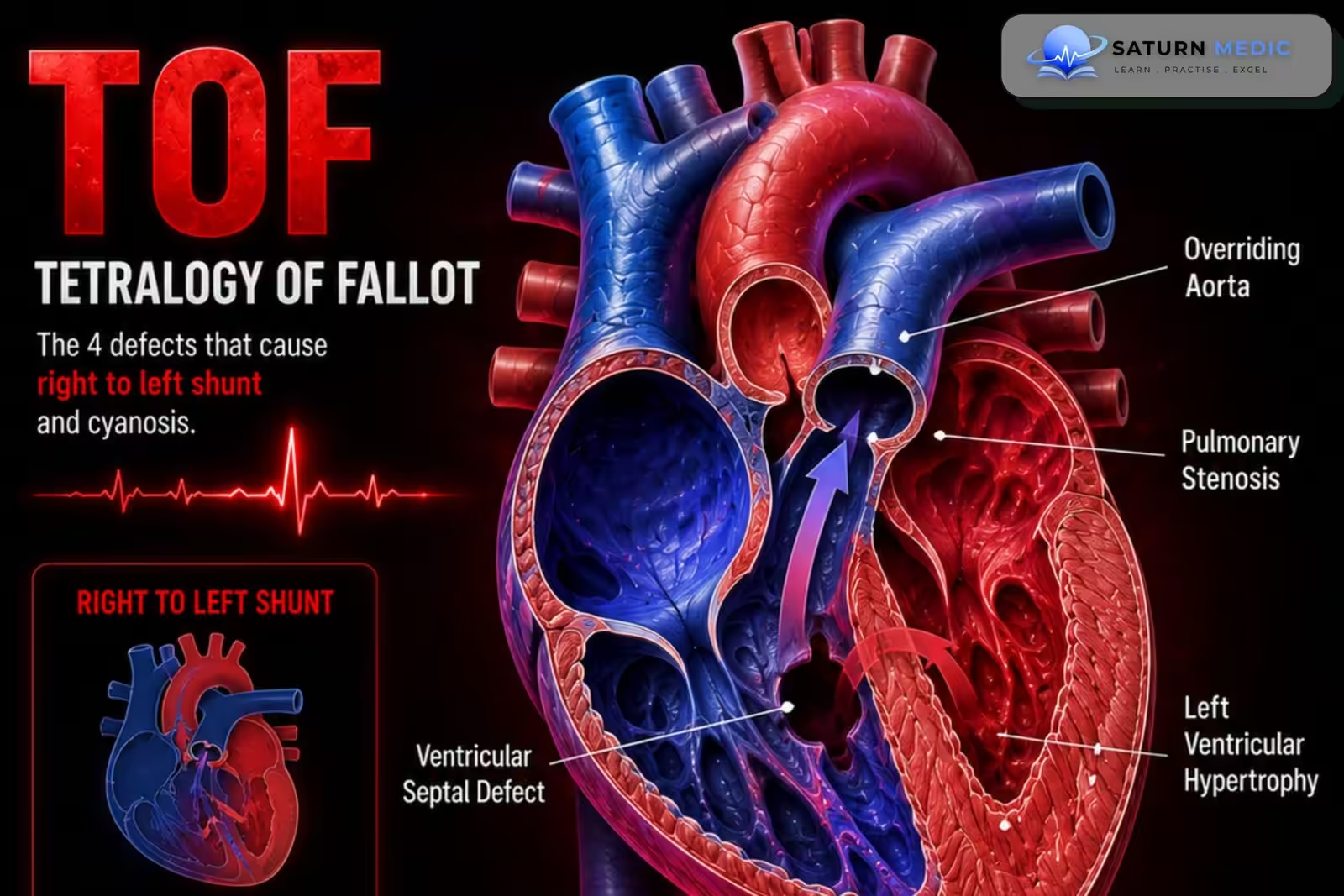
-Tetralogy of Fallot (TOF) is characterized by four congenital cardiac defects:
– Right ventricular outflow tract obstruction (RVOTO) due to pulmonary infundibular stenosis
– Right ventricular hypertrophy (RVH)
– Ventricular septal defect (VSD)
– Overriding aorta
Etiology & Risk Factors
- – Prematurity
– Maternal exposure during pregnancy:
– Rubella infection (first trimester)
– Alcohol consumption
– Phenytoin use (fetal hydantoin syndrome)
– Prostaglandin use
– Respiratory distress syndrome
– Trisomies (e.g., Down syndrome)
Pathophysiology
- During fetal development:
– Anterior and superior deviation of the infundibular septum causes: - Malaligned VSD
- Overriding aorta
- Right ventricular outflow tract obstruction (RVOT)
- Blood flow depends on the severity of RVOTO:
- Large VSD causes equal right and left ventricular pressures.
- Mild RVOTO → predominantly left-to-right shunt → minimal cyanosis.
- Severe RVOTO → right-to-left shunt → deoxygenated blood enters systemic circulation.
- Persistent right-to-left shunting eventually results in right ventricular hypertrophy
Clinical Presentation
- General
– Mild Tetralogy of Fallot (TOF) :
– May be asymptomatic initially.
– Symptoms of heart failure may develop after 4–6 weeks.
– Cyanosis:
– Severity depends on RVOTO.
– Mild obstruction:
– Minimal cyanosis (“Pink Tet”).
– Severe obstruction:
– Marked cyanosis present from birth.
Tet Spells (Hypercyanotic Spells)
– Peak incidence:
– 2–4 months of age.
– Triggered by:
– Crying
– Feeding
– Defecation
History Taking
Assess for:
– Cyanosis since birth
– Feeding difficulty
– Failure to thrive
– Dyspnea
– Hypercyanotic (tet) spells
– Episodes triggered by crying or feeding
– Squatting behavior during activity
– Exercise intolerance
– Symptoms of heart failure
Investigations
- Echocardiography (Confirmatory Test)
– Prenatal diagnosis is increasingly common with fetal echocardiography.
– Postnatal echocardiography confirms:
– Right ventricular outflow tract obstruction
– Ventricular septal defect
– Overriding aorta
– Right ventricular hypertrophy
– Measures RV outflow tract pressure gradient.
– Cardiac catheterization may supplement evaluation. - Chest X-ray
– Boot-shaped heart
– Upturned cardiac apex
– Right ventricular hypertrophy
Diagnosis
Diagnosis is confirmed by:
– Echocardiography.
Supportive investigations:
– Prenatal fetal echocardiography.
– Chest X-ray.
– ECG.
– Pulse oximetry.
– Hyperoxia test.
– Cardiac catheterization when required.
Management
1. Initial Management
Severe Right Ventricular Outflow Tract Obstruction (RVOTO)
Prostaglandin E₁ (PGE₁) infusion to keep the ductus arteriosus patent until definitive surgery.
2. Management of Acute Tet Spell (Hypercyanotic Spell)
Initial Measures
100% oxygen
Knee-to-chest position (or squatting in older children)
Morphine (or alternatives such as fentanyl, midazolam, or ketamine)
If No Improvement
IV sodium bicarbonate (to correct metabolic acidosis)
IV fluids (to improve right ventricular filling)
Phenylephrine (to increase systemic vascular resistance)
IV propranolol (to reduce sympathetic activity and relieve RV outflow tract spasm)
3. Heart Failure Management
Digoxin (inotropic support)
Loop diuretics (e.g., furosemide)
Avoid ACE inhibitors, as they may decrease systemic vascular resistance and precipitate tet spells.
4. Definitive Treatment
Complete Surgical Repair (Preferred)
Performed within the first year of life, typically before 6 months.
The procedure includes:
Patch closure of the ventricular septal defect (VSD)
Relief of the right ventricular outflow tract obstruction (RVOTO) by resecting obstructive infundibular muscle
5. Palliative Surgery
If complete repair cannot be performed initially:
Blalock–Thomas–Taussig (BT) shunt (most common)
Central shunt
Other palliative shunts when indicated
6. Long-Term Follow-Up
Regular cardiology follow-up
Monitor for:
Heart failure
Arrhythmias (e.g., ventricular tachycardia)
Neurodevelopmental complications
Complications
- Hypercyanotic (tet) spells
- Heart failure
- Cardiac arrhythmias (especially ventricular tachycardia)
- Neurodevelopmental impairment
- Chronic hypoxemia if untreated
- Death if severe disease is not corrected
Prognosis
- Without surgery: Approximately 50% of patients do not survive beyond the first 3 years of life.
- With corrective surgery: >90% of patients survive beyond 25 years of age.
- Lifelong cardiology follow-up is required to monitor for heart failure and arrhythmias.
Key Points / Clinical Pearls
- Tetralogy of Fallot (TOF) is the most common cyanotic congenital heart disease.
- In Tetralogy of Fallot (TOF) There are 4 defects: Pulmonary stenosis (RVOTO), VSD, Overriding aorta, RVH.
- Severity of cyanosis depends on the degree of RV outflow tract obstruction.
- Tet spells are life-threatening and are treated with oxygen, knee-to-chest position, and morphine.
- Squatting increases systemic vascular resistance (SVR) and decreases the right-to-left shunt, improving oxygenation.
- Gold-standard diagnostic test: Echocardiography.
- Definitive treatment: Complete surgical repair in infancy (usually before 6 months of age).
- Boot-shaped heart on chest X-ray is a classic finding.
- National Center for Biotechnology Information (NIH). Tetralogy of Fallot, StatPearls.
- 2025 ACC/AHA/HRS/ISACHD/SCAI Guideline for the Management of Adults With Congenital Heart Disease. Circulation. 2025. doi: 10.1161/CIR.0000000000001402.
- Bouzas B, Kilner PJ, Gatzoulis MA. Pulmonary Regurgitation: Not a Benign Lesion. Eur Heart J. 2005;26:433-439. PMID: 15640261.
- Geva T. Pulmonary Regurgitation After Tetralogy of Fallot Repair: Clinical Features, Sequelae, and Timing of Pulmonary Valve Replacement. Circ Cardiovasc Imaging. 2008;1:11-15. PMID: 18377431.
- MedlinePlus, National Library of Medicine (NIH). Tetralogy of Fallot: Medical Encyclopedia.