

Clinical Subject Page
Tricuspid Valve Disease (Stenosis & Regurgitation)
Also called
ICD-10
Specialty
Onset
Reviewed
-
OverviewOverview
-
Etiology & Risk FactorsEtiology & Risk Factors
-
PathophysiologyPathophysiology
-
Clinical PresentationClinical Presentation
-
History TakingHistory Taking
-
Tricuspid Stenosis Vs Tricuspid RegurgitationTricuspid Stenosis Vs Tricuspid Regurgitation
-
InvestigationsInvestigations
-
DiagnosisDiagnosis
-
ManagementManagement
-
ComplicationsComplications
-
PrognosisPrognosis
-
Key Points / Clinical PearlsKey Points / Clinical Pearls
Overview
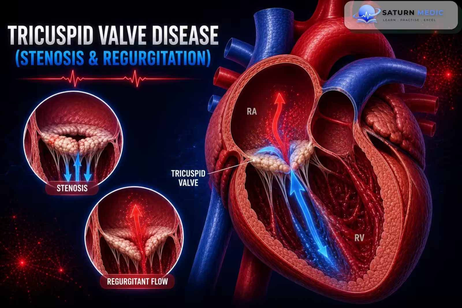
Tricuspid valve disease affects the tricuspid valve, which lies between the right atrium and right ventricle. It includes:
- Tricuspid stenosis (TS): Narrowing of the tricuspid valve, which reduces blood flow from the right atrium to the right ventricle. It is extremely rare.
- Tricuspid regurgitation (TR): Failure of the tricuspid valve to close properly, causing backflow of blood from the right ventricle into the right atrium during systole.
Etiology & Risk Factors
1. Tricuspid Stenosis (TS)
Most common causes:
Rheumatic fever (most common)
Infective endocarditis (especially in IV drug users)
2. Tricuspid Regurgitation (TR)
Common causes:
Right ventricular dilation (e.g., right-sided heart failure)
Pressure overload of the right ventricle
Pulmonary hypertension (e.g., cor pulmonale, secondary to mitral stenosis)
Left-sided heart failure
Infective endocarditis (especially in IV drug users)
Rheumatic fever
Carcinoid syndrome
Connective tissue disorders (e.g., Marfan syndrome)
General Risk Factors (for Tricuspid Valve Disease)
History of rheumatic fever
Infective endocarditis
Intravenous (IV) drug use
Pulmonary hypertension
Right-sided heart failure
Left-sided heart failure
Connective tissue disorders (e.g., Marfan syndrome)
Carcinoid syndrome
Pathophysiology
1. Tricuspid Stenosis (TS)
Narrowing of the tricuspid valve obstructs blood flow from the right atrium (RA) to the right ventricle (RV) during diastole.
This causes:
Increased right atrial pressure
Systemic venous congestion
Reduced RV filling
Decreased cardiac output
Eventually right-sided heart failure
Simple Flow:
Tricuspid valve narrowing → ↓ RA → RV blood flow → ↑ Right atrial pressure → Systemic venous congestion → Right-sided heart failure
2. Tricuspid Regurgitation (TR)
Incomplete closure of the tricuspid valve allows blood to flow back from the RV into the RA during systole.
This causes:
Right atrial volume overload
Right ventricular volume overload
Systemic venous congestion
Progressive right-sided heart failure
Simple Flow:
Tricuspid valve fails to close → Blood regurgitates into RA → RA & RV volume overload → Systemic venous congestion → Right-sided heart failure
Clinical Presentation
Tricuspid Stenosis (TS)
Symptoms
Fatigue
Exercise intolerance
Symptoms of right-sided heart failure
Leg swelling (peripheral edema)
Abdominal swelling (ascites)
Examination
Delayed diastolic murmur heard best at the left lower sternal border (4th left intercostal space).
May have a presystolic crescendo murmur.
Tricuspid Regurgitation (TR)
Symptoms
Fatigue
Exercise intolerance
Symptoms of right-sided heart failure
Peripheral edema
Ascites
Examination
Holosystolic murmur heard best at the left lower sternal border (4th left intercostal space).
Carvallo sign: the murmur becomes louder during inspiration.
History Taking
Symptoms
“Do you get short of breath?”
“Do you feel tired more easily than before?”
“Do you have chest pain?”
“Do you feel your heart racing or skipping beats (palpitations)?”
“Have you ever fainted or felt dizzy?”
Symptoms of Right-Sided Heart Failure
“Have you noticed swelling in your legs or ankles?”
“Has your abdomen become swollen?”
“Have you gained weight because of fluid retention?”
Risk Factors
“Have you ever had rheumatic fever?”
“Have you had infective endocarditis?”
“Do you have a history of heart failure or pulmonary hypertension?”
“Do you use intravenous (IV) drugs?”
“Have you ever been told you have a heart valve problem?”
Tricuspid Stenosis Vs Tricuspid Regurgitation
Tricuspid Stenosis vs Tricuspid Regurgitation
| Feature | Tricuspid Stenosis | Tricuspid Regurgitation |
|---|---|---|
| Murmur | Diastolic rumble at left lower sternal border, increases with inspiration (Rivero-Carvallo-like augmentation) | Holosystolic at left lower sternal border/subxiphoid, increases with inspiration (true Rivero-Carvallo sign) |
| JVP findings | Giant "a" wave (forceful RA contraction against obstruction); slow "y" descent | Prominent/giant "v" wave (CV wave); rapid "y" descent; may see systolic pulsation of liver |
| Pulse character | Normal arterial pulse; low cardiac output if severe | Normal; pulsatile liver, peripheral oedema common |
| Key echo finding | Diastolic pressure gradient across TV (mean gradient ≥5 mmHg suggests significant TS); restricted leaflet motion; doming | Color Doppler regurgitant jet; vena contracta, PISA/EROA, RA/RV dilation, systolic flow reversal in hepatic veins (severe) |
| Right heart chamber effect | RA dilation predominates; RV often normal/small initially | RA and RV dilation; progressive RV dysfunction |
| Common associations | Mitral stenosis (rheumatic), carcinoid syndrome | Pulmonary hypertension, left heart failure, atrial fibrillation, pacemaker/ICD leads |
| Definitive treatment | Balloon valvuloplasty (if pliable, non-calcified) or surgical repair/replacement | Treat underlying cause (PHTN, LV disease); diuretics for symptoms; surgical repair/replacement or transcatheter repair if severe/symptomatic |
Investigations
1. Transthoracic Echocardiography (TTE) – Gold Standard
First-line investigation
Assesses:
Valve anatomy and morphology
Severity of tricuspid stenosis (TS) or tricuspid regurgitation (TR)
Cardiac structure and hemodynamics
2. ECG
Assesses:
Cardiac rhythm
Signs of ventricular hypertrophy
3. Chest X-ray
Evaluates:
Pulmonary congestion
Other lung pathology
4. Advanced Investigations (if needed)
Transesophageal echocardiography (TEE) – better assessment of valve anatomy and complications
Cardiac MRI – assesses structural heart disease
Cardiac catheterization – evaluates valve hemodynamics and cardiac pressures
Exercise stress testing – if symptoms are unclear
Diagnosis
Diagnostic Approach
Suspect TS or TR based on:
Clinical symptoms
Characteristic heart murmur on examination
Confirm the diagnosis with Transthoracic Echocardiography (TTE).
Physical Examination Findings
Tricuspid Stenosis (TS):
Delayed diastolic murmur at the left lower sternal border (4th left intercostal space)
Tricuspid Regurgitation (TR):
Holosystolic murmur
Carvallo sign: murmur becomes louder with inspiration
Management
1. Medical (Supportive) Management
Treat the underlying cause (e.g., heart failure, pulmonary hypertension, infective endocarditis).
Manage heart failure symptoms.
Monitor disease progression with:
Regular history and physical examination
Periodic transthoracic echocardiography (TTE)
Consider endocarditis prophylaxis when indicated.
Secondary prevention of rheumatic fever if appropriate.
Anticoagulation if indicated (e.g., after prosthetic valve replacement).
2. Interventional/Surgical Management
Patients with severe and/or symptomatic valve disease should be referred for intervention.
Valve Repair
Valve reconstruction (annuloplasty) to restore valve function.
Valvuloplasty (balloon or surgical commissurotomy) for valvular stenosis.
Valve Replacement
Surgical valve replacement if repair is not feasible.
Choice of mechanical or bioprosthetic valve depends on:
Age
Comorbidities
Ability to take long-term anticoagulation
Patient preference
3. Acute Decompensation
Patients presenting with:
Acute heart failure
Cardiogenic shock
Severe arrhythmias
require:
Urgent stabilization
Immediate cardiology consultation
Consideration of urgent valve intervention
Complications
-
- Right-sided heart failure
- Progressive systemic venous congestion
- Cardiac arrhythmias
- Infective endocarditis
- Worsening valve dysfunction leading to decompensated heart failure
Prognosis
- Prognosis depends on:
- Severity of the valve disease
- Underlying cause
- Development of heart failure
- Timely medical or surgical treatment
- Patients with mild disease may remain stable with regular follow-up.
- Severe symptomatic disease has a poorer prognosis if left untreated but improves with appropriate valve repair or replacement.
Key Points / Clinical Pearls
-
- Tricuspid stenosis (TS) is rare and is most commonly caused by rheumatic fever.
- Tricuspid regurgitation (TR) is more common and is often due to right ventricular dilation or pulmonary hypertension.
- Both conditions can lead to right-sided heart failure.
- Gold-standard investigation: Transthoracic echocardiography (TTE).
- TS murmur: Delayed diastolic murmur.
- TR murmur: Holosystolic murmur that increases with inspiration (Carvallo sign).
- Treat the underlying cause, manage heart failure symptoms, and consider valve repair or replacement for severe symptomatic disease.
- National Center for Biotechnology Information (NIH). Tricuspid Stenosis, StatPearls.
- Otto CM, Nishimura RA, Bonow RO, et al. 2020 ACC/AHA Guideline for the Management of Patients With Valvular Heart Disease. Circulation. 2021;143:e72-e227. PMID: 33972115.
- National Center for Biotechnology Information (NIH). Tricuspid Valve Repair, StatPearls.
- Sorajja P, Whisenant B, Hamid N, et al; TRILUMINATE Pivotal Investigators. Transcatheter Repair for Patients With Tricuspid Regurgitation. N Engl J Med. 2023;388:1833-1842. PMID: 36876753.
- MedlinePlus, National Library of Medicine (NIH). Tricuspid Valve Regurgitation: Medical Encyclopedia.