

Clinical Subject Page
Ventricular Septal Defect (VSD)
ICD-10
Specialty
Onset
Reviewed
-
OverviewOverview
-
Etiology & Risk FactorsEtiology & Risk Factors
-
PathophysiologyPathophysiology
-
Clinical PresentationClinical Presentation
-
History TakingHistory Taking
-
InvestigationsInvestigations
-
DiagnosisDiagnosis
-
ManagementManagement
-
ComplicationsComplications
-
PrognosisPrognosis
-
Key Points / Clinical PearlsKey Points / Clinical Pearls
Overview
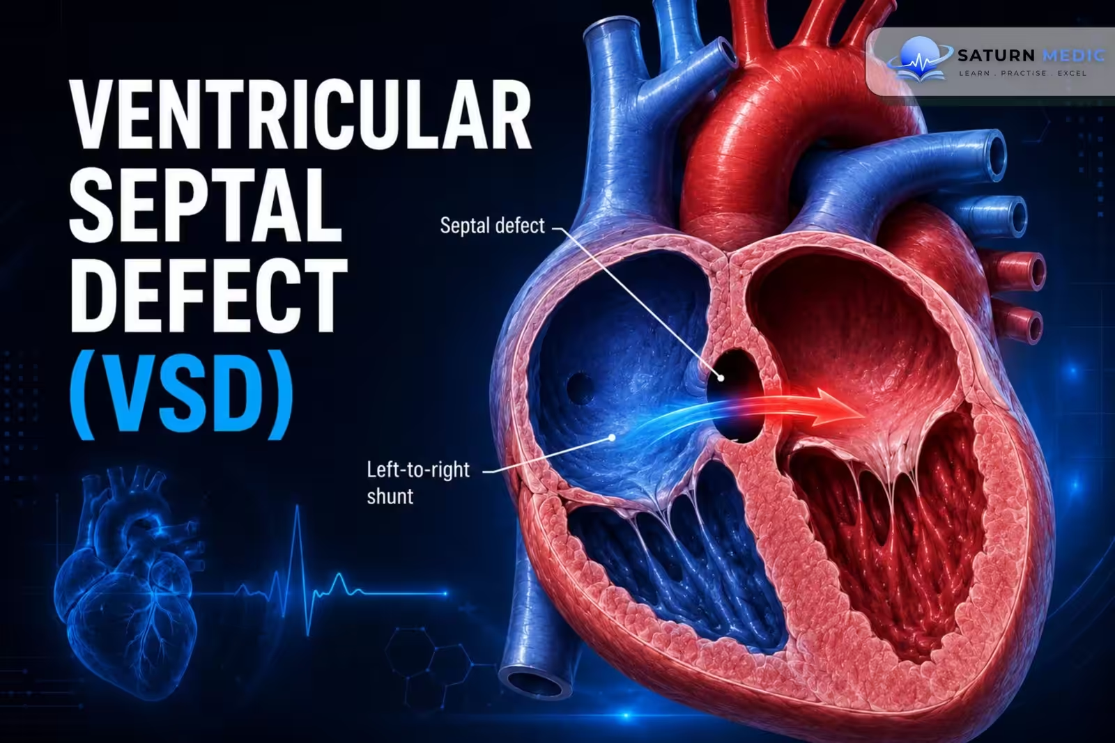
Ventricular Septal Defect (VSD) is An abnormal communication between the left and right ventricle that results in a left-to-right shunt
Etiology & Risk Factors
- – Genetic syndromes
– Down syndrome (most common)
– Edwards syndrome
– Patau syndrome
– Cri-du-chat syndrome
– Apert syndrome
– Intrauterine infections (e.g., TORCH)
– Maternal risk factors
– Diabetes
– Obesity
– Smoking
– Rare acquired causes
– Post-myocardial infarction
– Aortic valve replacement
Pathophysiology
- – Most Ventricular Septal Defect (VSD) occur in the membranous part of the ventricular septum.
– Defect causes a left-to-right shunt leading to:
– Right ventricular volume overload → RV eccentric hypertrophy
– Increased pulmonary blood flow
– Increased pulmonary artery pressure → pulmonary hypertension
– Decreased cardiac output
– Left ventricular volume overload → LV eccentric hypertrophy
– Increased oxygen saturation in the right ventricle and pulmonary artery
– Long-standing pulmonary hypertension may progress to Eisenmenger syndrome.
Clinical Presentation
- Small Ventricular Septal Defect (VSD)
– Usually asymptomatic.
Medium or Large Ventricular Septal Defect (VSD)
– Heart failure develops during the first weeks to months of life.
– Symptoms appear after pulmonary vascular resistance decreases after birth.
– Hyperdynamic precordium may be present.
Auscultation
– Harsh holosystolic murmur at the left lower sternal border.
– Murmur becomes louder with handgrip.
– Murmur is typically louder in small defects.
– Systolic thrill in the 3rd or 4th left intercostal space.
– Mid-diastolic murmur at the cardiac apex.
– Loud pulmonic S2 if pulmonary hypertension develops
History Taking
Assess for:
– Feeding difficulty
– Poor weight gain or failure to thrive
– Tachypnea
– Sweating during feeding
– Fatigue
– Recurrent respiratory infections
– Symptoms of heart failure
– Exercise intolerance in older children
– History suggestive of pulmonary hypertension
Investigations
Echocardiography (Confirmatory Test)
– Transthoracic echocardiography (TTE) is preferred.
Assess:
– Defect size
– Shunt volume
– Qp:Qs ratio
– Pulmonary artery pressure
– Associated cardiac abnormalities
– Doppler echocardiography is especially useful for detecting small VSDs.
– TEE may be required for detailed anatomy or intraoperative guidance.
ECG
Small defects:
– Often normal.
Medium/Large defects:
– Left atrial enlargement (P mitrale)
– Left ventricular hypertrophy
– Increased QRS amplitude
– Left axis deviation
– If pulmonary hypertension develops:
– Right ventricular hypertrophy
– Right axis deviation
– P pulmonale
– PR prolongation
– Right bundle branch block
Diagnosis
Diagnosis is confirmed by:
-Transthoracic echocardiography with Doppler.
Supportive investigations:
– ECG
– Chest X-ray
– Cardiac MRI/CT
– Cardiac catheterization when indicated
Management
Small Asymptomatic Ventricular Septal Defect (VSD)
– Spontaneous closure is common.
– Surgery is rarely required.
– Regular follow-up echocardiography.
Symptomatic or Large Ventricular Septal Defect (VSD)
Surgical repair:
– Patch closure
– Alternative transcatheter closure in selected patients.
Indications for Surgery
– Infants with large left-to-right shunts and symptoms
– Failure to thrive
Complications
- Arrhythmias
- Heart failure
- Eisenmenger syndrome
- Infective endocarditis
- Aortic regurgitation
Prognosis
- – Small Ventricular Septal Defect (VSD) frequently close spontaneously.
– Prognosis is excellent after successful repair.
– Untreated large defects may progress to pulmonary hypertension and Eisenmenger syndrome.
– Ventricular Septal Defect (VSD) closure is contraindicated in Eisenmenger syndrome.
Key Points / Clinical Pearls
-
Ventricular Septal Defect (VSD) is the most common congenital heart defect.
A left-to-right shunt is the hallmark of Ventricular Septal Defect (VSD).
Small Ventricular Septal Defect (VSD) are often asymptomatic and may close spontaneously during childhood.
Large Ventricular Septal Defect (VSD) can cause heart failure, pulmonary hypertension, and poor growth.
A harsh holosystolic murmur at the lower left sternal border is the classic auscultatory finding.
Echocardiography is the diagnostic test of choice.
Pulmonary hypertension may progress to Eisenmenger syndrome if left untreated.
Medical therapy treats heart failure symptoms but does not close the defect.
Surgical or transcatheter closure is indicated for hemodynamically significant Ventricular Septal Defect (VSD).
Early diagnosis and timely repair prevent irreversible pulmonary vascular disease and improve long-term outcomes.
- Alahmadi MH, Oliver TI. National Center for Biotechnology Information (NIH). Ventricular Septal Defect, StatPearls.
- 2025 ACC/AHA/HRS/ISACHD/SCAI Guideline for the Management of Adults With Congenital Heart Disease. Circulation. 2025. doi: 10.1161/CIR.0000000000001402.
- Brown KN, Adnan G, Kanmanthareddy A. National Center for Biotechnology Information (NIH). Catheter Management of Ventricular Septal Defect, StatPearls.
- MedlinePlus, National Library of Medicine (NIH). Ventricular Septal Defect: Medical Encyclopedia.
- Basit H, Wallen TJ, Sergent BN. National Center for Biotechnology Information (NIH). Eisenmenger Syndrome, StatPearls.