

Clinical Subject Page
Addison’s Disease
Addison’s Disease is primary adrenal insufficiency caused by destruction or dysfunction of the adrenal cortex, resulting in inadequate production of cortisol and aldosterone.
Also called
ICD-10
Specialty
Onset
Reviewed
-
OverviewOverview
-
Etiology & Risk FactorsEtiology & Risk Factors
-
PathophysiologyPathophysiology
-
Clinical PresentationClinical Presentation
-
History TakingHistory Taking
-
Physical ExaminationPhysical Examination
-
InvestigationsInvestigations
-
DiagnosisDiagnosis
-
ManagementManagement
-
ComplicationsComplications
-
PrognosisPrognosis
-
Key Points / Clinical PearlsKey Points / Clinical Pearls
Overview
- Addison’s Disease occurs when the adrenal cortex cannot produce sufficient glucocorticoids and mineralocorticoids.
- The most common cause in many settings is autoimmune adrenalitis. Other causes include tuberculosis, adrenal hemorrhage, metastatic disease, and certain infections.
-Typical features include:
- Fatigue
- Weight loss
- Hyperpigmentation
- Hypotension
- Hyponatremia
- Hyperkalemia
- Hypoglycemia
Etiology & Risk Factors
–Etiology of Addison’s Disease:
-Autoimmune
The most common cause of Addison’s Disease in developed countries is autoimmune destruction of the adrenal cortex.
Autoantibodies against 21-hydroxylase are commonly present.
-Infectious
- Tuberculosis
- HIV-associated infections
-Other Causes
- Bilateral adrenal hemorrhage
- Adrenal metastases
- Adrenal infiltration
- Adrenalectomy
-Risk Factors for Addison’s Disease:
- Autoimmune disease
- Family history of autoimmune disease
- Previous tuberculosis
- HIV infection
- Adrenal surgery
- Adrenal hemorrhage
- Metastatic malignancy
Pathophysiology
Adrenal cortex destruction → ↓ cortisol + ↓ aldosterone → ↓ cortisol-mediated glucose regulation + ↓ vascular responsiveness + renal sodium loss → hypotension + hyponatremia + dehydration + hyperkalemia → ↑ ACTH due to loss of negative feedback → increased melanocortin activity → hyperpigmentation
Clinical Presentation
-Symptoms:
- Fatigue
- Generalized weakness
- Weight loss
- Loss of appetite
- Nausea
- Vomiting
- Abdominal pain
- Diarrhea
- Salt craving
-Signs:
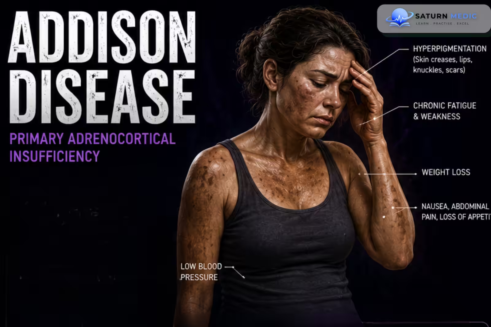
- Hyperpigmentation
- Hypotension
- Vitiligo in autoimmune disease
- Darkening of palmar creases
- Darkening of scars
- Pigmentation of oral mucosa
History Taking
-Ask about:
- Fatigue and weakness
- Weight loss
- Appetite
- Nausea and vomiting
- Abdominal pain
- Diarrhea
- Salt craving
- Dizziness
- Postural symptoms
- Episodes of fainting
- Hyperpigmentation
- Hypoglycemic symptoms
Physical Examination
-General Examination:
- Weight
- Blood pressure
- Orthostatic blood pressure
- Heart rate
-Look for:
- Weight loss
- Dehydration
- Hyperpigmentation
-System-Specific Examination:
–Skin
Look for:
- Generalized hyperpigmentation
- Palmar crease pigmentation
- Knuckle pigmentation
- Scar pigmentation
–Endocrine
- Assess for evidence of other autoimmune endocrine diseases, particularly:
- Thyroid disease
- Type 1 Diabetes Mellitus
Investigations
-Biochemistry / Specific Tests:
- Typical findings include:
- Low serum sodium
- High serum potassium
- Low glucose in some patients
- Increased urea
- Increased creatinine during dehydration
- Low serum bicarbonate in severe cases
-Serum Cortisol:
- A low morning serum cortisol strongly supports adrenal insufficiency.
- ACTH
- High ACTH → primary adrenal insufficiency
- Low or inappropriately normal ACTH → suggests secondary/tertiary adrenal insufficiency
-Special / Confirmatory Tests:
-ACTH Stimulation Test
The 250-µg cosyntropin stimulation test is commonly used to confirm adrenal insufficiency.
In primary adrenal insufficiency:
ACTH administration → inadequate rise in serum cortisol
-Adrenal Autoantibodies
21-hydroxylase antibodies support an autoimmune cause
Diagnosis
Diagnosis of Addison’s Disease is established by demonstrating inadequate cortisol production and determining whether the problem is primary or central.
-Typical findings in Addison Disease:
- Low morning cortisol
- High ACTH
- Inadequate cortisol response to ACTH stimulation
- Low aldosterone
- High renin
- Hyponatremia
- Hyperkalemia
Management
1. First-Line / Emergency Management
Suspected Addisonian Crisis
Immediate treatment:
- IV hydrocortisone
- Rapid intravenous isotonic fluid replacement
- Correction of hypoglycemia
- Electrolyte monitoring
- Treatment of the precipitating cause
Common precipitants include:
- Infection
- Surgery
- Trauma
- Vomiting or inability to take medication
2. Definitive Treatment
Lifelong hormone replacement is usually required.
The goals are:
- Replace cortisol
- Replace aldosterone
- Prevent adrenal crisis
- Educate the patient about stress dosing
3. Medical Treatment
Glucocorticoid Replacement
Common options include:
- Hydrocortisone
- Prednisolone
Hydrocortisone is commonly used because its pharmacological profile resembles physiological cortisol secretion.
4. Supportive Management
- Patient education
- Stress-dose steroid instructions
- Medical alert identification
- Emergency injectable hydrocortisone when appropriate
Complications
- Addisonian crisis
- Hypotension
- Shock
- Severe dehydration
- Hyponatremia
- Hyperkalemia
- Hypoglycemia
- Acute kidney injury
- Cardiac arrhythmias due to severe electrolyte abnormalities
Prognosis
–With appropriate lifelong hormone replacement and patient education, the prognosis is generally good.
-The major life-threatening complication is adrenal crisis, which can occur when glucocorticoid requirements increase and replacement is inadequate.
-Prognosis is improved by:
- Correct diagnosis
- Adequate hormone replacement
- Proper stress dosing
- Emergency steroid access
- Patient education
Key Points / Clinical Pearls
- Addison’s Disease is primary adrenal insufficiency caused by failure of the adrenal cortex.
- It causes ↓ cortisol and usually ↓ aldosterone.
- Autoimmune adrenalitis is the most common cause in many settings.
- Classic features include fatigue, weight loss, hypotension, and hyperpigmentation.
- Salt craving may occur due to mineralocorticoid deficiency.
- Typical laboratory findings are hyponatremia, hyperkalemia, and sometimes hypoglycemia.
- Low cortisol + high ACTH suggests primary adrenal insufficiency.
- High renin + low aldosterone indicates mineralocorticoid deficiency.
- 21-hydroxylase antibodies support an autoimmune cause.
- The ACTH stimulation test is commonly used to confirm adrenal insufficiency.
- Hyperpigmentation helps distinguish primary from secondary adrenal insufficiency.
- Treatment requires lifelong glucocorticoid replacement
- Bornstein SR, Allolio B, Arlt W, et al. Diagnosis and Treatment of Primary Adrenal Insufficiency: An Endocrine Society Clinical Practice Guideline. J Clin Endocrinol Metab. 2016;101(2):364-389. Journal of Clinical Endocrinology & Metabolism .
- Husebye ES, Pearce SH, Krone NP, Kämpe O. Adrenal Insufficiency. Lancet. 2021;397(10274):613-629. The Lancet .
- Betterle C, Dal Pra C, Mantero F, Zanchetta R. Autoimmune Adrenal Insufficiency and Autoimmune Polyendocrine Syndromes: Autoantibodies, Autoantigens, and Their Applicability in Diagnosis and Disease Prediction. Endocr Rev. 2002;23(3):327-364.
- National Institute of Diabetes and Digestive and Kidney Diseases (NIDDK). Addison's Disease . National Institutes of Health.
- National Library of Medicine (NIH). Addison Disease . StatPearls.