

Clinical Subject Page
Pheochromocytoma
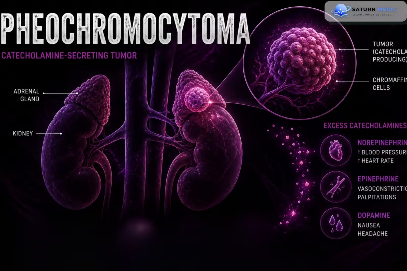
Pheochromocytoma is a catecholamine-producing tumor arising from chromaffin cells, usually in the adrenal medulla. It causes excessive secretion of epinephrine, norepinephrine, or both, often resulting in episodic or persistent hypertension.
Also called
ICD-10
Specialty
Onset
Reviewed
-
OverviewOverview
-
Etiology & Risk FactorsEtiology & Risk Factors
-
PathophysiologyPathophysiology
-
Clinical PresentationClinical Presentation
-
History TakingHistory Taking
-
Physical ExaminationPhysical Examination
-
InvestigationsInvestigations
-
DiagnosisDiagnosis
-
ManagementManagement
-
ComplicationsComplications
-
PrognosisPrognosis
-
Key Points / Clinical PearlsKey Points / Clinical Pearls
Overview
–Pheochromocytoma is a rare neuroendocrine tumor that produces excessive catecholamines.
-Classic clinical features include:
-
-
- Episodic headache
- Sweating
- Palpitations
- Hypertension
- The classic triad is highly suggestive but is not present in every patient.
-
Etiology & Risk Factors
-Etiology:
-Pheochromocytoma arises from chromaffin cells of the adrenal medulla.
Catecholamine secretion may include:
- Norepinephrine
- Epinephrine
- Dopamine
Most tumors are located in the adrenal glands, while related extra-adrenal tumors are called paragangliomas.
-A significant proportion of pheochromocytomas are associated with hereditary syndromes or pathogenic germline variants.
-Important associations include:
- Multiple Endocrine Neoplasia type 2 (MEN2)
- Von Hippel-Lindau disease (VHL)
- Neurofibromatosis type 1 (NF1)
-Risk Factors:
- Family history of pheochromocytoma or paraganglioma
- MEN2
- Von Hippel-Lindau disease
- Neurofibromatosis type 1
- Hereditary paraganglioma syndromes
Pathophysiology
Chromaffin cell tumor → excessive catecholamine production → α-adrenergic stimulation → vasoconstriction → hypertension
Catecholamine excess → β-adrenergic stimulation → tachycardia + increased cardiac contractility → palpitations
Catecholamine surges → episodic autonomic activation → headache + sweating + palpitations + hypertension
Chronic catecholamine excess → cardiovascular stress → arrhythmias + cardiomyopathy + myocardial injury
Clinical Presentation
-Symptoms:
- Episodic headache
- Excessive sweating
- Palpitations
- Anxiety
- Tremor
- Chest discomfort
- Shortness of breath
- Abdominal pain
- Nausea
- Weight loss
-Signs:
- Hypertension
- Tachycardia
- Orthostatic hypotension
- Pallor
- Tremor
- Excessive sweating
- Weight loss
- Hyperglycemia
History Taking
-Ask about:
- Headaches
- Episodic sweating
- Palpitations
- Blood pressure episodes
- Chest pain
- Shortness of breath
- Tremor
- Anxiety or panic-like episodes
- Weight loss
- Abdominal pain
- Constipation
- Heat intolerance
- Orthostatic dizziness
- Previous hypertensive crises
Physical Examination
–General Examination
- Blood pressure
- Heart rate
- Orthostatic blood pressure
- Body weight
- Hydration status
-Look for:
- Hypertension
- Tachycardia
- Pallor
- Sweating
- Tremor
- Weight loss
-System-Specific Examination:
–Cardiovascular
-Assess for:
- Hypertension
- Tachycardia
- Arrhythmias
- Cardiomyopathy
- Heart failure
–Neurological
-Assess for:
- Headache
- Neurological deficits
- Evidence of previous cerebrovascular complications
–Genetic/Syndromic Examination
-Look for features suggesting:
- Neurofibromatosis type 1
- MEN2
- Von Hippel-Lindau disease
Investigations
-Biochemistry / Specific Tests:
-Plasma Free Metanephrines
A highly sensitive screening test.
-Measure:
- Metanephrine
- Normetanephrine
-24 Hour Urinary Fractionated Metanephrines
An alternative biochemical test.
May also measure:
- Urinary catecholamines
-Additional tests may include:
- Glucose
- Electrolytes
- Renal function
- Liver function
–Imaging
After biochemical evidence of catecholamine excess, localization is usually performed with:
CT or MRI
- Adrenal CT
- Adrenal MRI
- Abdominal/pelvic imaging when extra-adrenal disease is suspected
Diagnosis
-Diagnosis is based on :
Diagnosis requires evidence of excess catecholamine production together with appropriate clinical and/or radiological findings.
-Initial biochemical evaluation
- Plasma free metanephrines
- or
- 24-hour urinary fractionated metanephrines
Markedly elevated results strongly support the diagnosis.
Management
1. First-Line / Emergency Management
-Suspected Pheochromocytoma Crisis
This is a medical emergency.
Management may include:
- Immediate cardiovascular stabilization
- Alpha-adrenergic blockade
- Intravenous fluids when appropriate
- Treatment of severe hypertension
- Management of arrhythmias
- Intensive monitoring
If a patient is not already adequately alpha-blocked, pure beta-blockade should not be started first, because unopposed alpha-adrenergic stimulation can worsen hypertension.
2. Definitive Treatment
Surgical removal of the tumor is the definitive treatment for localized resectable pheochromocytoma.
3. Medical Treatment
Alpha-Adrenergic Blockade
Common options include:
- Phenoxybenzamine
- Selective α1-blockers such as doxazosin
Used preoperatively to:
- Control blood pressure
- Reduce catecholamine-related complications
4. Surgical / Procedural Treatment
- Adrenalectomy
- Usually performed laparoscopically for appropriately selected localized tumors.
- Open surgery may be considered for:
-Before surgery:
- Alpha blockade → volume optimization → beta blockade if needed → tumor resectio
Complications
- Hypertensive crisis
- Cardiac arrhythmias
- Catecholamine-induced cardiomyopathy
- Acute heart failure
- Myocardial ischemia/injury
- Stroke
- Pulmonary edema
- Acute kidney injury
- Hyperglycemia
Prognosis
-The prognosis is generally good for localized tumors that are completely resected.
-Prognosis depends on:
- Tumor location
- Tumor size
- Genetic background
- Completeness of resection
- Presence of metastatic disease
- Recurrence
Key Points / Clinical Pearls
- Pheochromocytoma is a catecholamine-producing tumor of chromaffin cells, usually in the adrenal medulla.
- The classic symptoms are headache, sweating, and palpitations.
- Hypertension may be episodic or persistent.
- Patients may have normal blood pressure between episodes.
- Important hereditary associations include MEN2, von Hippel-Lindau disease, NF1, and hereditary paraganglioma-pheochromocytoma syndromes.
- Plasma free metanephrines or urinary fractionated metanephrines are key biochemical tests.
- Biochemical testing generally comes before localization imaging.
- CT or MRI is used to localize the tumor after biochemical evidence is established.
- Genetic evaluation should be considered in many patients with pheochromocytoma.
- Lenders JWM, Duh QY, Eisenhofer G, et al. Pheochromocytoma and Paraganglioma: An Endocrine Society Clinical Practice Guideline. J Clin Endocrinol Metab. 2014;99(6):1915-1942. Journal of Clinical Endocrinology & Metabolism .
- Taïeb D, Hicks RJ, Hindie E, et al. European Association of Nuclear Medicine Practice Guideline for Radionuclide Imaging of Pheochromocytoma and Paraganglioma. Eur J Nucl Med Mol Imaging. 2012;39(12):1977-1989.
- Neumann HPH, Young WF Jr, Eng C. Pheochromocytoma and Paraganglioma. N Engl J Med. 2019;381(6):552-565. New England Journal of Medicine .
- Eisenhofer G, Lenders JWM, Siegert G, et al. Plasma Methoxytyramine: A Novel Biomarker of the Metastatic Potential of Pheochromocytoma and Paraganglioma. J Clin Endocrinol Metab. 2012;97(8):2790-2799.
- National Cancer Institute (NIH). Pheochromocytoma and Paraganglioma .
- National Library of Medicine (NIH). Pheochromocytoma . StatPearls.