

Clinical Subject Page
Lung Abscess
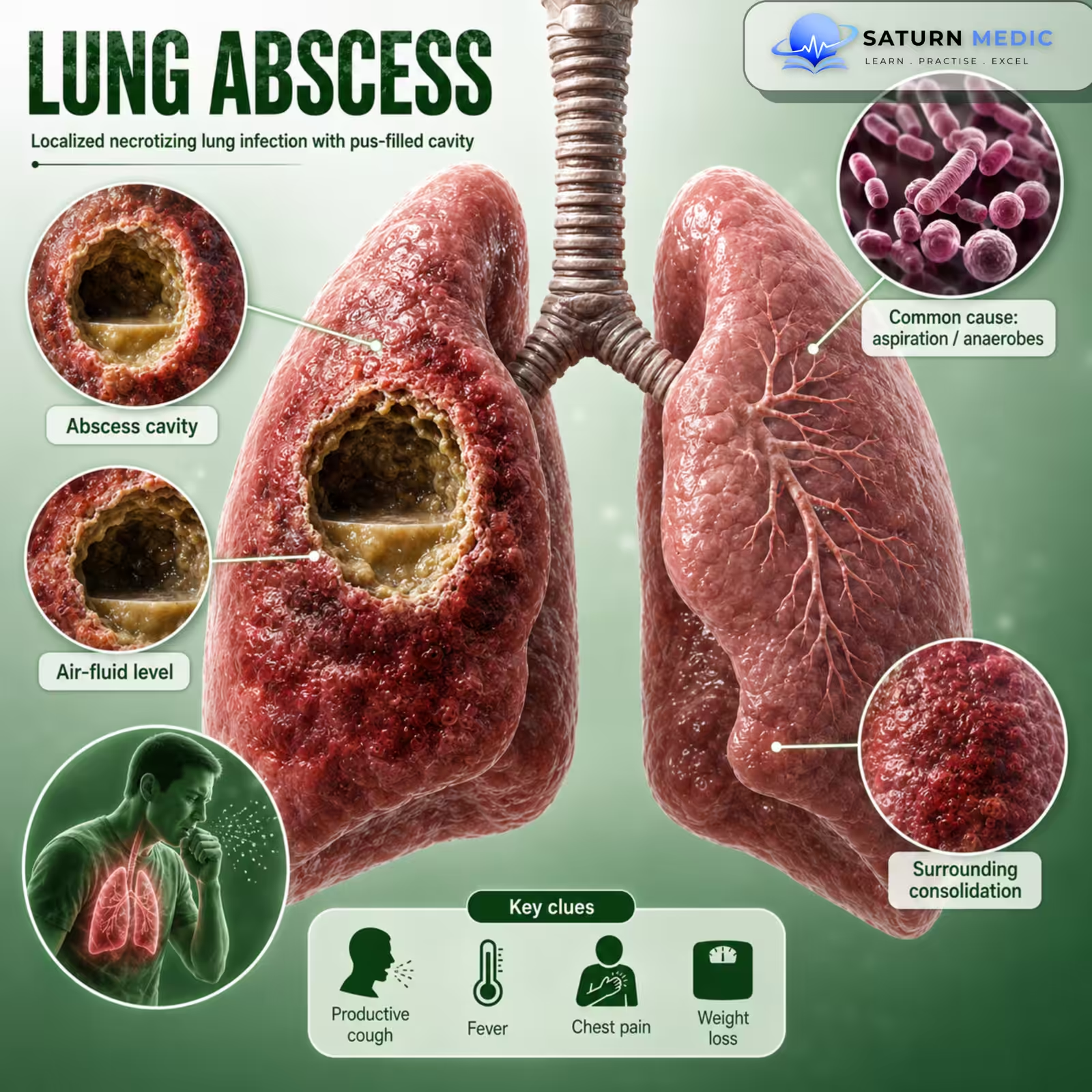
A lung abscess is a localized collection of pus inside the lung tissue caused by an infection. It develops when part of the lung becomes infected, leading to tissue destruction and the formation of a cavity filled with pus.
Also called
ICD-10
Specialty
Onset
Reviewed
-
OverviewOverview
-
Etiology & Risk FactorsEtiology & Risk Factors
-
PathophysiologyPathophysiology
-
Clinical PresentationClinical Presentation
-
History TakingHistory Taking
-
Physical ExaminationPhysical Examination
-
InvestigationsInvestigations
-
DiagnosisDiagnosis
-
ManagementManagement
-
ComplicationsComplications
-
PrognosisPrognosis
-
Key Points / Clinical PearlsKey Points / Clinical Pearls
Overview
- Most common cause: Aspiration of mouth or throat secretions into the lungs.
- Common organisms: Mainly anaerobic bacteria, but aerobic bacteria can also cause it.
Etiology & Risk Factors
-Etiology
- The main causes of a lung abscess are:
- Aspiration of oropharyngeal secretions (most common cause)
- Bronchial obstruction (e.g., lung cancer or a foreign body)
- Pneumonia or bronchiectasis
- Impaired mucus clearance (e.g., cystic fibrosis)
- Immunocompromised state
- Lung Abscess
-Risk Factors
- Impaired consciousness (e.g., alcohol or drug intoxication, stroke) leading to aspiration
- Difficulty swallowing due to neurological disorders or vocal cord paralysis
- Poor oral hygiene, periodontal disease, dental abscesses, or tonsillitis
- Bronchial obstruction (lung tumor, foreign body)
- Immunocompromised patients
- Chronic lung diseases such as bronchiectasis or cystic fibrosis
Pathophysiology
Aspiration of oropharyngeal secretions (most common) → Bacteria enter the lung → Localized lung infection (suppurative inflammation) → Lung tissue necrosis (destruction) → Formation of a pus-filled cavity (lung abscess) → Cough with foul-smelling sputum, fever, and cavitary lesion with an air–fluid level on imaging
Other causes: Bronchial obstruction / hematogenous spread / contiguous spread / necrotizing pneumonia → Localized infection → Tissue necrosis → Lung abscess
Clinical Presentation
- Fever
- Productive cough with foul-smelling, purulent sputum (classic feature)
- Night sweats
- Hemoptysis (coughing up blood)
- Pleuritic chest pain
- Weight loss, anorexia, and fatigue
- Symptoms usually develop gradually over weeks to months (may also be acute).
History Taking
- Do you have a fever?
- Do you have a cough?
- Are you coughing up foul-smelling or pus-like sputum? (Most important)
- Have you coughed up blood (hemoptysis)?
- Do you have chest pain when taking a deep breath?
- Have you had choking episodes or difficulty swallowing? (Aspiration risk)
- Do you drink alcohol heavily or have episodes of loss of consciousness? (Aspiration risk)
- Have you had pneumonia recently?
- Have you had weight loss or night sweats?
- Do you have poor dental hygiene or a recent dental infection?
Physical Examination
-vital signs:
- Fever
- tachycardia
-Respiratory Examination :
- Inspect: Digital clubbing (in chronic cases)
- Percussion: Dullness over the affected area
- Auscultation: Amphoric breath sounds may be heard over the abscess
-Key Physical Signs :
- Fever
- Digital clubbing (chronic lung abscess)
- Dullness to percussion
- Amphoric breath sounds
- The most important physical findings are fever, dullness to percussion, and amphoric breath sounds; digital clubbing may be present in chronic disease.
Investigations
-Chest X-ray – Initial test
- CT chest with IV contrast – Gold standard
- Shows thick-walled cavity with an air–fluid level
-Laboratory Tests
- CBC – ↑ WBC (leukocytosis)
- Blood cultures
- Sputum Gram stain and culture (including aerobic and anaerobic cultures)
-Additional Tests (if indicated)
- Bronchoscopy – Suspected tumor or foreign body
- Echocardiography – If infective endocarditis is suspected
- Swallow assessment – If aspiration is suspected
- HIV test – If immunocompromised or no obvious risk factor
Diagnosis
-Diagnosis is based on:
- History: Fever, cough with foul-smelling purulent sputum, aspiration risk
- Imaging:
- Chest X-ray or
- CT chest with IV contrast (gold standard)
- Classic finding: Thick-walled lung cavity with an air–fluid level
- Laboratory tests:
- CBC: Elevated WBC
- Blood cultures
- Sputum Gram stain and culture
Related Topics
Management of Lung abscess
- Admit the patient.
- Obtain blood and sputum cultures, then start empiric antibiotics immediately.
- First-line antibiotics (anaerobic coverage):
- Ampicillin–sulbactam (preferred)
- Clindamycin (alternative)
- Duration: 3–6 weeks or until clinical and radiologic improvement.
- Tailor antibiotics according to culture results.
- Drainage (bronchoscopic or percutaneous) if:
- Large abscess (> 6–8 cm)
- Significant hemoptysis
- No response to antibiotics
- Surgery is rarely required for refractory cases.
- Treat the underlying cause (e.g., aspiration, foreign body, bronchial obstruction, dysphagia).
Complications of Lung abscess
- Pleural empyema
- Pleural effusion
- Bronchopulmonary fistula
- Pneumothorax (from rupture into the pleural cavity)
- Massive hemoptysis
- Recurrence of the abscess
- Chronic bronchiectasis or pneumatoceles
- The most serious complications are rupture into the pleural cavity (causing empyema or pneumothorax) and massive hemoptysis.
Prognosis of Lung abscess
- Generally good with early diagnosis and appropriate antibiotic treatment.
- Most patients improve with prolonged antibiotic therapy (3–6 weeks).
- Prognosis is worse if:
- Treatment is delayed.
- The abscess is large or does not respond to antibiotics.
- Serious complications develop (e.g., empyema, pneumothorax, massive hemoptysis).
- The underlying cause is not treated.
Key Points / Clinical Pearls of Lung abscess
- Lung abscess = A pus-filled cavity in the lung caused by infection.
- Most common cause: Aspiration of oropharyngeal secretions.
- Most common organisms: Anaerobic bacteria.
- Classic symptoms: Fever, cough with foul-smelling purulent sputum, hemoptysis, chest pain.
- Diagnosis: Chest X-ray and CT chest (gold standard) showing a thick-walled cavity with an air–fluid level.
- Treatment: Start empiric antibiotics with anaerobic coverage after obtaining cultures; continue for 3–6 weeks. Drainage or surgery may be needed if the abscess is large or does not respond.
- Complications: Pleural empyema, pleural effusion, pneumothorax, bronchopulmonary fistula, massive hemoptysis.
- Prognosis: Usually good with early diagnosis and appropriate treatment.
- Sabbula BR, Rammohan G, Sharma S, Akella J. National Center for Biotechnology Information (NIH). Lung Abscess, StatPearls.
- MedlinePlus, National Library of Medicine (NIH). Lung Abscess: Medical Encyclopedia.
- Jain V, Vashisht R, Yilmaz G, Bhardwaj A. National Center for Biotechnology Information (NIH). Pneumonia Pathology, StatPearls.
- Kelogrigoris M, Tsagouli P, Stathopoulos K, et al. Percutaneous Drainage of Lung Abscesses: Outcomes and Factors Affecting the Successful Outcome. J Thorac Dis. 2019. PMID: 31463117.
- Shebl E, Paul M. National Center for Biotechnology Information (NIH). Parapneumonic Pleural Effusions and Empyema Thoracis, StatPearls.