

Clinical Subject Page
Pleural Empyema
Pleural Empyema is the accumulation of pus within the pleural cavity. It is an infected exudative pleural effusion that most commonly develops as a complication of pneumonia and requires prompt antibiotic therapy and pleural drainage.
Also called
ICD-10
Specialty
Onset
Reviewed
-
OverviewOverview
-
Etiology & Risk FactorsEtiology & Risk Factors
-
PathophysiologyPathophysiology
-
Clinical PresentationClinical Presentation
-
History TakingHistory Taking
-
Physical ExaminationPhysical Examination
-
InvestigationsInvestigations
-
DiagnosisDiagnosis
-
ManagementManagement
-
ComplicationsComplications
-
PrognosisPrognosis
-
Key Points / Clinical PearlsKey Points / Clinical Pearls
Overview
Pleural Empyema results from bacterial infection of the pleural space, leading to purulent
pleural fluid. Disease progression occurs through exudative, fibrinopurulent, and
organizing stages, with increasing pleural fibrosis and restriction of lung expansion if
untreated.
Etiology & Risk Factors
Etiology
Most common cause:
- Complication of bacterial pneumonia (parapneumonic infection)
Other causes:
- Thoracic surgery
- Chest trauma
- Esophageal rupture
- Lung abscess
- Bronchopleural fistula
- Spread from nearby infection
- Infected pleural procedure or chest tube
Common Organisms
- Streptococcus species
- Staphylococcus aureus
- Anaerobic bacteria
- Gram-negative bacteria
Risk Factors
- Pneumonia
- Aspiration
- Poor dental hygiene
- Alcohol misuse
- Diabetes mellitus
- Immunosuppression
- Chronic lung disease
- Recent thoracic surgery or trauma
- Delayed or inadequate treatment of pneumonia
Pathophysiology
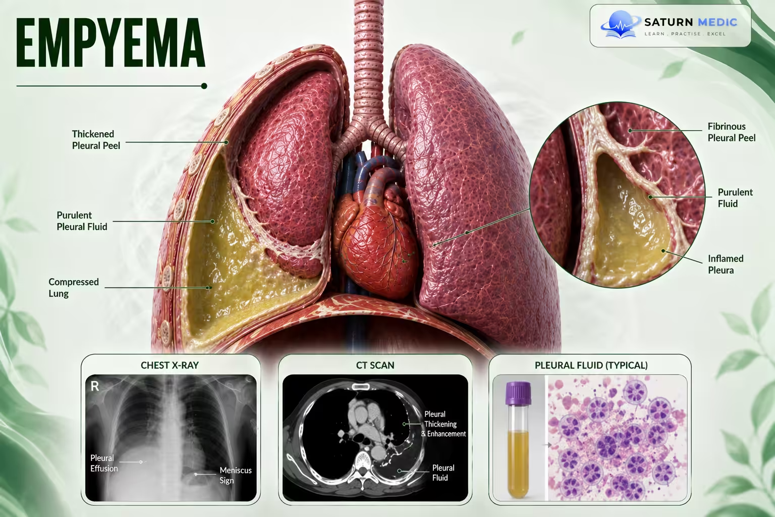
Pleural Empyema progresses through three stages:
Stage I (Exudative)
• Accumulation of fluid and pus
Stage II (Fibrinopurulent)
• Fibrin deposition
• Formation of septations and loculated pockets
Stage III (Organizing)
• Formation of thick fibrous pleural peel
• Restriction of lung movement (trapped lung)
Clinical Presentation
Symptoms
- Fever
- Chills
- Pleuritic chest pain
- Dyspnea
- Cough
- Purulent sputum
- Fatigue
- Malaise
- Night sweats
- Weight loss in chronic cases
Signs
- Fever
- Tachypnea
- Tachycardia
- Reduced oxygen saturation
- Reduced chest expansion on the affected side
- Dullness to percussion
- Reduced breath sounds
- Reduced vocal fremitus
Severe Disease
- Respiratory distress
- Hypoxia
- Hypotension
- Confusion
- Sepsis or septic shock
History Taking
-Assess for:
• Recent pneumonia
• Fever and chills
• Persistent cough
• Chest discomfort
• History of thoracic trauma
• Lung abscess
• Esophageal injury
• Previous hemothorax
Physical Examination
-Assess:
• Respiratory status
• Fever
• Signs of pleural infection
• Hemodynamic stability
Investigations
-Imaging
Chest X-ray
May show:
• Pleural opacity
• Meniscus sign
• Lenticular (biconvex) fluid collection
• Adjacent lung consolidation
Thoracic ultrasound
• Heterogeneous pleural fluid collection
Chest CT
May demonstrate:
• Split pleura sign
• Septations
• Loculated collections
• Compression of the adjacent lung
Pleural fluid analysis
Typical findings:
• Exudative effusion
• Grossly purulent appearance
• Positive Gram stain
• Positive bacterial culture
• pH <7.2
• Low glucose (<30–60 mg/dL)
• Elevated LDH
Diagnosis
-Diagnosis is based on:
• Clinical features
• Chest imaging
• Diagnostic thoracentesis
• Purulent pleural fluid
• Positive microbiological studies when present
Related Topics
Management of Pleural Empyema
Empiric antibiotic therapy
-Community-acquired empyema
• Ceftriaxone (or another second-/third-generation cephalosporin) PLUS:
• Metronidazole, or
• Clindamycin
OR
• Ampicillin-sulbactam
Hospital-acquired empyema
Provide MRSA and Pseudomonas coverage:
• Vancomycin PLUS one of:
• Cefepime + metronidazole
• Piperacillin-tazobactam
• Meropenem (if ESBL organisms are suspected)
-Additional points
• Tailor antibiotics according to pleural fluid culture.
• Aminoglycosides are not recommended due to poor pleural penetration.
• Intrapleural antibiotics are not recommended.
• Drainage should be performed as soon as possible.
Definitive management
Stage I
• Chest tube (thoracostomy)
• Consider intrapleural fibrinolytic therapy
Stage II
• Chest tube drainage
• VATS debridement if drainage is ineffective
Stage III
• VATS debridement
• Pleurectomy and lung decortication via open thoracotomy when indicated
Complications of Pleural Empyemara
• Recurrece
• Tension pneumothorax
• Cardiorespiratory compromise
Prognosis of Pleural Empyema
-Prognosis depends on:
• Early diagnosis
• Prompt pleural drainage
• Adequate antibiotic therapy
• Stage of disease at presentation
Early-stage empyema often responds well to chest tube drainage, whereas advanced
disease may require surgical intervention.
Key Points / Clinical Pearls of Pleural Empyema
• Most commonly follows pneumonia.
• Symptoms include fever, chills, cough, and chest discomfort.
• Pleural fluid is purulent, with low pH, low glucose, and elevated LDH.
• CT may show the split pleura sign.
• Management requires antibiotics plus drainage.
• Chest tube is first-line treatment.
• VATS or thoracotomy may be required in advanced disease.
- Iguina MM, Sharma S, Danckers M. National Center for Biotechnology Information (NIH). Thoracic Empyema, StatPearls.
- Shebl E, Paul M. National Center for Biotechnology Information (NIH). Parapneumonic Pleural Effusions and Empyema Thoracis, StatPearls.
- MedlinePlus, National Library of Medicine (NIH). Empyema: Medical Encyclopedia.
- Semenkovich TR, Olsen MA, Puri V, et al. Current State of Empyema Management. Ann Thorac Surg. 2018. PMC5964038.
- Kumar A, Sharma S. National Center for Biotechnology Information (NIH). Lung Decortication, StatPearls.