

Clinical Subject Page
Interstitial lung disease (ILD)
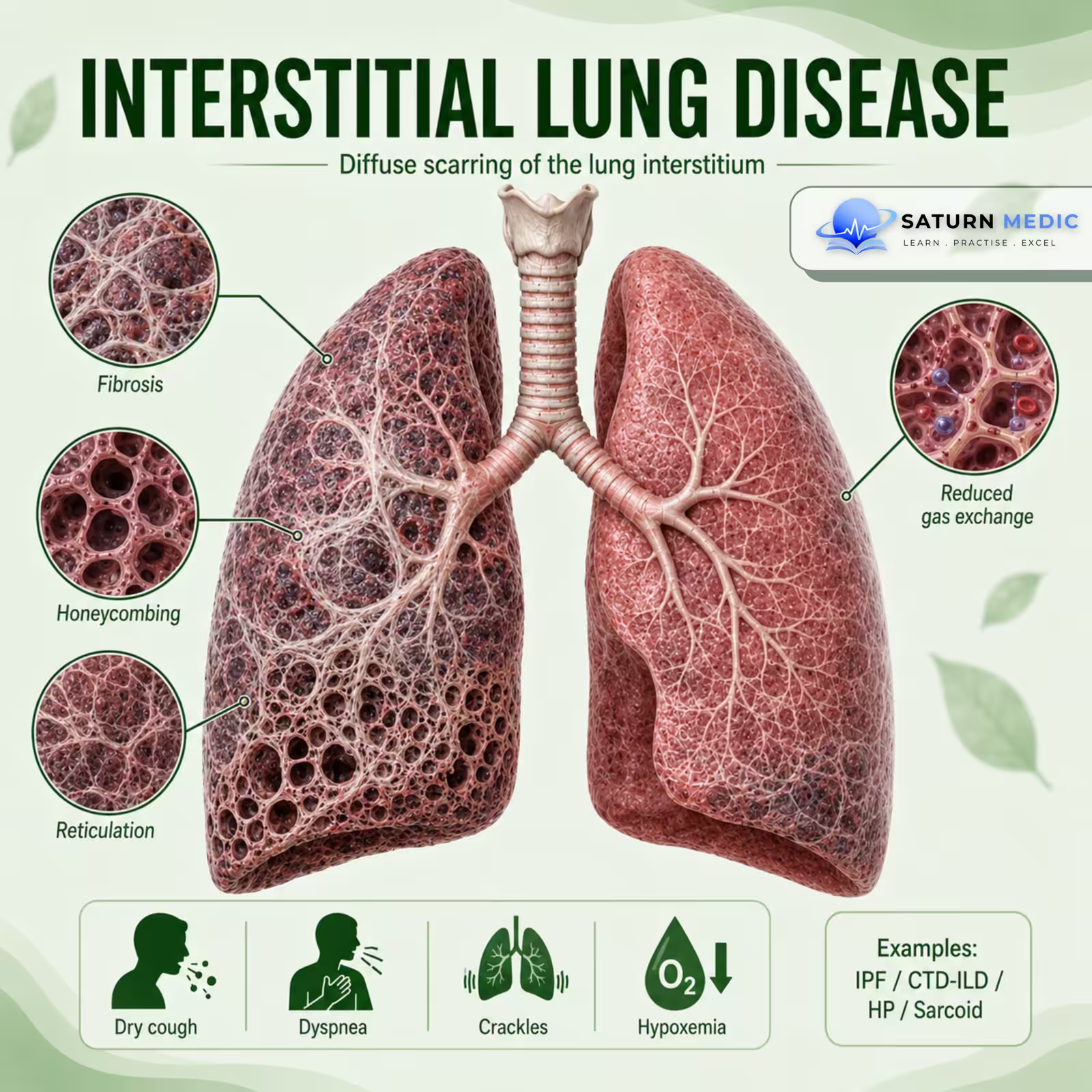
Interstitial lung disease (ILD) is a group of disorders that cause inflammation and/or scarring (fibrosis) of the lung interstitium, leading to stiff lungs and impaired oxygen transfer.
Also called
ICD-10
Specialty
Onset
Reviewed
-
OverviewOverview
-
Etiology & Risk FactorsEtiology & Risk Factors
-
PathophysiologyPathophysiology
-
Clinical PresentationClinical Presentation
-
History TakingHistory Taking
-
Physical ExaminationPhysical Examination
-
ClassificationClassification
-
InvestigationsInvestigations
-
DiagnosisDiagnosis
-
ManagementManagement
-
ComplicationsComplications
-
PrognosisPrognosis
-
Key Points / Clinical PearlsKey Points / Clinical Pearls
Overview
Interstitial lung disease (ILD) includes many different diseases that affect the:
- Lung interstitium
- Alveoli
- Small airways
- Pulmonary blood vessels
Persistent injury may cause:
Inflammation → Fibrosis → Stiff lungs → Reduced oxygen transfer
Patients typically present with progressive exertional dyspnea and a dry cough.
Etiology & Risk Factors
Common Causes
Idiopathic
-
Idiopathic pulmonary fibrosis (IPF)
-
Other idiopathic interstitial pneumonias
Autoimmune Diseases
-
Rheumatoid arthritis
-
Systemic sclerosis
-
Inflammatory myopathies
-
Sjögren syndrome
-
Systemic lupus erythematosus
Occupational & Environmental Exposure
-
Asbestos
-
Silica
-
Coal dust
-
Organic dusts
-
Bird exposure
-
Mold exposure
Drugs
-
Amiodarone
-
Methotrexate
-
Bleomycin
-
Nitrofurantoin
Other Causes For Interstitial lung disease (ILD)
-
Sarcoidosis
-
Hypersensitivity pneumonitis
-
Radiation therapy
-Risk Factors
-
Increasing age
-
Cigarette smoking
-
Occupational dust exposure
-
Bird or mold exposure
-
Autoimmune disease
-
Family history of Interstitial lung disease (ILD)
-
Use of pulmonary-toxic medications
Pathophysiology
Lung injury or abnormal immune response
⬇
Inflammation of the alveoli and interstitium
⬇
Repeated tissue injury
⬇
Abnormal healing response
⬇
Fibroblast activation + Collagen deposition
⬇
Interstitial fibrosis
⬇
Thickened alveolar-capillary membrane
⬇
Stiff lungs + Impaired oxygen diffusion
⬇
Restrictive lung disease + Hypoxemia
Key Concept
Fibrosis reduces lung compliance, making the lungs difficult to expand.
Clinical Presentation
Symptoms
-
Progressive exertional dyspnea
-
Persistent dry cough
-
Fatigue
-
Reduced exercise tolerance
-
Chest discomfort
Advanced Disease
-
Dyspnea at rest
-
Weight loss
-
Cyanosis
Signs
-
Fine inspiratory crackles
-
Digital clubbing
-
Tachypnea
-
Reduced oxygen saturation
Advanced Signs of Interstitial lung disease (ILD)
-
-
Cyanosis
-
Signs of pulmonary hypertension
-
Signs of right-sided heart failure
-
History Taking
-In Interstitial lung disease (ILD), Ask about:
-
- Shortness of breath?
- Is it progressive?
- Dry cough?
- When did symptoms start?
- Occupational exposure?
- Dust exposure?
- Asbestos or silica exposure?
- Bird exposure?
- Mold exposure?
- Smoking history?
- Current and previous medications?
- Previous radiation therapy?
Physical Examination
-General Examinaion
Look for:
-
Tachypnea
-
Cyanosis
-
Reduced oxygen saturation
-
Digital clubbing
Chest Examination
-
Fine end-inspiratory crackles
-
Reduced chest expansion in advanced disease
Look for an Underlying Cause for Interstitial lung disease (ILD)
-
Joint swelling or deformity
-
Skin thickening
-
Skin rash
-
Muscle weakness
-
Raynaud phenomenon
Advanced Interstitial lung disease (ILD)
Look for:
-
-
Raised JVP
-
Peripheral edema
-
Signs of pulmonary hypertension
-
Signs of cor pulmonale
-
Classification
Interstitial Lung Disease · Classification — ATS/ERS 2013
Investigations
High-Resolution CT (HRCT) Chest — Key Imaging Test
May show:
-
Ground-glass opacities
-
Reticulation
-
Traction bronchiectasis
-
Honeycombing
The pattern and distribution help identify the specific type of ILD.
Pulmonary Function Tests
Typically show:
-
Restrictive pattern
-
Reduced total lung capacity
-
Reduced diffusion capacity (DLCO)
Oxygen Assessment
-
Pulse oximetry
-
Exercise oxygen assessment
-
Arterial blood gas in severe disease
Blood Tests
Depending on the suspected cause:
-
CBC
-
Inflammatory markers
-
Autoimmune serology
Additional Tests
When needed:
-
-
Bronchoscopy with bronchoalveolar lavage
-
Lung biopsy
-
Multidisciplinary review
-
Diagnosis
Interstitial Lung Disease (ILD) · Diagnostic Criteria
| ILD Category | Key Examples | Typical HRCT Pattern |
|---|---|---|
| Idiopathic Interstitial Pneumonias (IIPs) | IPF NSIP COP DIP, RB-ILD, AIP, LIP | UIP (honeycombing + traction bronchiectasis, basal/subpleural) for IPF; ground-glass + subpleural sparing for NSIP; consolidation + ground-glass for COP |
| Connective Tissue Disease-ILD | RA-ILD, SSc-ILD, myositis-ILD (anti-MDA5/Jo-1), SLE-ILD, Sjogren's-ILD | NSIP pattern most common; UIP in RA; organising pneumonia in myositis; specific patterns vary by CTD |
| Hypersensitivity Pneumonitis (HP) | Farmer's lung (thermophilic actinomycetes), Bird fancier's lung (avian proteins), hot tub lung, chemical exposures | Acute/subacute: ground-glass + mosaic attenuation + air trapping. Chronic: fibrotic HP with UIP-like features; head-cheese sign |
| Granulomatous ILD | Sarcoidosis, hypersensitivity pneumonitis, berylliosis | Sarcoidosis: perilymphatic nodules, upper/mid lobe, bilateral hilar lymphadenopathy |
| Drug-Induced ILD | Amiodarone, methotrexate, nitrofurantoin, bleomycin, checkpoint inhibitors, biologics | Variable — organising pneumonia, NSIP, or diffuse alveolar damage depending on drug and mechanism |
| Occupational / Environmental ILD | Asbestosis, silicosis, coal worker's pneumoconiosis, berylliosis | Asbestosis: UIP-like, bilateral pleural plaques; silicosis: upper lobe nodules, eggshell calcification of nodes |
| Other / Rare ILDs | LAM (lymphangioleiomyomatosis), pulmonary Langerhans cell histiocytosis, eosinophilic pneumonia, PAP (alveolar proteinosis) | LAM: diffuse thin-walled cysts in young women; PLCH: upper lobe cysts + nodules in smokers; PAP: crazy paving |
Related Topics
Management
Interstitial Lung Disease (ILD) · Treatment by Type
| ILD Type | Treatment of Choice | Key Notes |
|---|---|---|
| IPF (Idiopathic Pulmonary Fibrosis) | Nintedanib or Pirfenidone | Antifibrotics — not immunosuppressants. Both slow FVC decline by ~50%. Do NOT use corticosteroids (harmful — PANTHER trial). Treat gastro-oesophageal reflux (common comorbidity, worsens IPF). Early transplant referral. |
| NSIP (Non-Specific Interstitial Pneumonia) | Prednisolone ± Mycophenolate or Azathioprine | Generally steroid-responsive, especially cellular NSIP. Fibrotic NSIP responds less well. Steroid-sparing agent added for maintenance. Monitor for CTD underlying cause. |
| COP (Cryptogenic Organising Pneumonia) | Prednisolone (0.75–1 mg/kg/day) | Highly corticosteroid-responsive — dramatic improvement usually within weeks. Prolonged taper over 6–12 months to prevent relapse (common if stopped too early). Excellent prognosis. |
| Hypersensitivity Pneumonitis (HP) | Antigen removal + Prednisolone (acute/subacute) | Most important step is complete antigen avoidance — disease may resolve without drugs if done early. Corticosteroids speed resolution in acute/subacute HP. Chronic fibrotic HP: antigen avoidance + consider nintedanib (progressive fibrotic HP). Relapses occur with re-exposure. |
| CTD-ILD (RA, SSc, Myositis) | Mycophenolate mofetil or Azathioprine ± steroids | Treat underlying CTD + ILD simultaneously. SSc-ILD: mycophenolate first-line (SLS II trial); nintedanib also approved. Myositis-ILD (MDA5+): often aggressive — high-dose steroids + calcineurin inhibitor + rituximab. RA-ILD: methotrexate may worsen ILD — discontinue and switch. |
| Sarcoidosis (pulmonary) | Prednisolone if symptomatic/progressive | Stage I/II often remits spontaneously — observe first. Treat if symptomatic, progressive, or extrapulmonary involvement (cardiac, CNS, hypercalcaemia). Steroid-sparing: methotrexate, azathioprine, hydroxychloroquine. Refractory: infliximab. |
| Drug-Induced ILD | Stop the offending drug + Prednisolone if severe | Most cases improve or resolve on drug withdrawal. Corticosteroids for severe/rapidly progressive cases (e.g. checkpoint inhibitor pneumonitis — high-dose IV methylprednisolone). Some drug-ILDs are irreversible (bleomycin, nitrofurantoin fibrosis). |
| Asbestosis / Silicosis (Pneumoconioses) | No disease-modifying therapy — supportive care only | Remove from exposure immediately. Treat complications (infection, respiratory failure, cor pulmonale). Asbestosis: annual surveillance for mesothelioma/lung cancer (high risk). Silicosis: whole lung lavage for silicoproteinosis; GM-CSF for autoimmune PAP if coexistent. |
| DIP / RB-ILD (Smoking-Related ILD) | Smoking cessation + Prednisolone if persistent | DIP and RB-ILD are almost exclusively smokers. Smoking cessation is the primary and often sufficient treatment. Corticosteroids if residual disease after cessation. Good prognosis if smoking stopped early. |
| Acute Exacerbation of IPF / AIP | High-dose IV methylprednisolone + ICU-level supportive care | Acute exacerbation of IPF: in-hospital mortality >50%. High-dose steroids empirically (evidence limited). Ventilatory support; consider NIV/HFNO over invasive ventilation. Urgent transplant activation if listed. AIP (Hamman-Rich): fulminant DAMP — steroids, ICU, high mortality. |
| Progressive Pulmonary Fibrosis (PPF) — non-IPF | Nintedanib | INBUILD trial: nintedanib slows FVC decline in non-IPF fibrotic ILDs that are progressing despite optimal treatment (HP, CTD-ILD, unclassifiable ILD). A major paradigm shift — antifibrotic use now extends beyond IPF. |
Complications
- Progressive pulmonary fibrosis
- Chronic hypoxemia
- Respiratory failure
- Pulmonary hypertension
- Cor pulmonale
- Acute exacerbation
- Increased risk of lung cancer
- Death
Prognosis
- Prognosis varies greatly depending on the specific Interstitial lung disease (ILD) .
- Some inflammatory Interstitial lung disease (ILD) may improve with treatment.
- Fibrotic Interstitial lung disease (ILD) are often progressive.
- Prognosis is worse with:
- Extensive fibrosis
- Declining lung function
- Severe hypoxemia
- Pulmonary hypertension
- Acute exacerbations
- Early diagnosis and treatment of reversible causes may improve outcomes.
Key Points / Clinical Pearls
- Interstitial lung disease (ILD) is a group of diseases, not a single disease.
- Progressive dyspnea and dry cough are classic symptoms.
- Fine inspiratory crackles are a common examination finding.
- HRCT is the key imaging test for Interstitial lung disease (ILD).
- Pulmonary function tests usually show a restrictive pattern with reduced DLCO.
- Always ask about occupational, environmental, medication, and autoimmune causes.
- Honeycombing suggests established pulmonary fibrosis.
- Treatment depends on the specific Interstitial lung disease (ILD) subtype.
- Antoine MH, Mlika M. National Center for Biotechnology Information (NIH). Interstitial Lung Disease, StatPearls.
- Raghu G, Remy-Jardin M, Richeldi L, et al. Idiopathic Pulmonary Fibrosis (an Update) and Progressive Pulmonary Fibrosis in Adults: An Official ATS/ERS/JRS/ALAT Clinical Practice Guideline. Am J Respir Crit Care Med. 2022. PMID: 35486072.
- National Center for Biotechnology Information (NIH). Interstitial Pulmonary Fibrosis, StatPearls.
- MedlinePlus, National Library of Medicine (NIH). Interstitial Lung Disease: Medical Encyclopedia.
- National Center for Biotechnology Information (NIH). Restrictive Lung Disease, StatPearls.