

Saturn Medic

Clinical Subject Page

Bronchiectasis

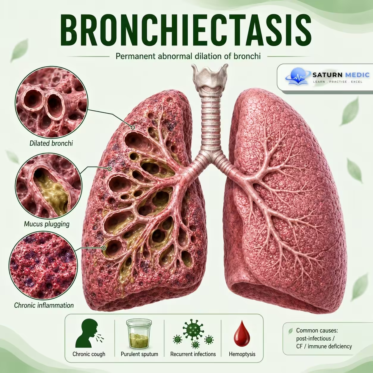
Bronchiectasis is a chronic lung disease characterized by permanent abnormal widening of the bronchi, leading to impaired mucus clearance, recurrent infections, and chronic airway inflammation.

Also called

Chronic suppurative airway disease

ICD-10

J47.9

Specialty

Pulmonology

Onset

Chronic

Reviewed

July 2026

On This Page

Overview

Damaged and widened bronchi cannot clear mucus effectively. Mucus accumulates, promoting recurrent infection and further airway damage.

Etiology & Risk Factors

Common Causes

  • Previous severe lung infection
  • Cystic fibrosis
  • Primary ciliary dyskinesia
  • Immune deficiency
  • Allergic bronchopulmonary aspergillosis (ABPA)
  • Airway obstruction
  • Autoimmune disease
  • Aspiration

Important Note

In many patients, no specific cause is identified (idiopathic bronchiectasis).

Risk Factors

  • Recurrent respiratory infections
  • Previous tuberculosis
  • Immunodeficiency
  • Chronic aspiration
  • Severe childhood lung infections
  • Underlying genetic disorders

Pathophysiology

Flow Chart:

Initial airway injury

Impaired mucus clearance

Mucus accumulation

Bacterial infection

Chronic inflammation

Airway wall destruction

Permanent bronchial dilatation

Further mucus retention and recurrent infection

Key Concept

This forms a vicious cycle of infection, inflammation, and airway damage.

Clinical Presentation

  1. Symptoms

    • Chronic productive cough
    • Daily sputum production
    • Recurrent chest infections
    • Dyspnea
    • Wheezing
    • Hemoptysis
    • Fatigue

    Sputum

    May be:

    • Large in volume
    • Purulent
    • Foul-smelling during infection

    Signs

    • Coarse crackles
    • Wheezing
    • Digital clubbing in advanced disease
    • Reduced oxygen saturation in severe disease
History Taking

  • Ask about:

    • Chronic cough?
    • Daily sputum?
    • Sputum amount and color?
    • Recurrent chest infections?
    • Hemoptysis?
    • Dyspnea or wheezing?
    • Previous tuberculosis or severe pneumonia?
    • Frequent childhood infections?
    • Recurrent sinus or ear infections?
    • Aspiration symptoms?

Physical Examination

  • Look for:

    • Respiratory distress
    • Cyanosis
    • Digital clubbing
    • Reduced oxygen saturation

    Chest Examination

    • Coarse inspiratory crackles
    • Wheezing
    • Rhonchi
    • Reduced breath sounds in advanced disease

Investigations

  • High-Resolution CT (HRCT) Chest — Key Test

    May show:

    • Dilated bronchi
    • Lack of normal bronchial tapering
    • Bronchi larger than accompanying pulmonary arteries
    • Bronchial wall thickening

    Sputum Tests

    • Sputum culture
    • Antibiotic sensitivity
    • Mycobacterial testing when indicated

    Pulmonary Function Tests

    May show:

    • Obstructive pattern
    • Mixed pattern
    • Sometimes normal results

    Blood Tests

    • CBC
    • Immunoglobulin levels
    • Tests for ABPA
    • Autoimmune testing when indicated

    Additional Tests

    Depending on the suspected cause:

    • Cystic fibrosis testing
    • Ciliary function testing
    • Bronchoscopy

Diagnosis

Bronchiectasis · Stepwise Diagnostic Approach — ERS 2017

1
Clinical history
Symptoms & risk factors
2
Physical examination
Signs & findings
3
HRCT thorax
Confirm diagnosis
4
Baseline investigations
Severity & function
5
Aetiology workup
Find the underlying cause
Step 1 — Clinical History
Cardinal symptoms
Chronic productive cough — most common and defining symptom; daily, often copious
Sputum may be mucoid, mucopurulent, or purulent. Volume >30 mL/day suggests significant disease.
Recurrent respiratory infections — ≥2 chest infections per year requiring antibiotics
Haemoptysis — blood-streaked sputum to massive haemoptysis; may be sole presentation
Dyspnea — progressive; correlates with extent of disease and FEV₁ decline
Fatigue, malaise, weight loss — especially during exacerbations
Pleuritic chest pain — if pleural involvement or pneumonia
Risk factors & red flags
Childhood pneumonia, measles, pertussis, or TB — most common antecedent causes
Recurrent sinusitis or otitis media — suggests PCD or immunodeficiency
Situs inversus + bronchiectasis — Kartagener syndrome (PCD subtype)
Infertility — PCD (male) or CF (both sexes)
Family history of lung disease — CF, PCD, alpha-1 antitrypsin deficiency
GI symptoms — malabsorption, steatorrhoea (CF); GERD (aspiration)
Rheumatoid arthritis, inflammatory bowel disease, connective tissue disorders
Symptoms >3 months duration without alternative explanation = investigate for bronchiectasis
Step 2 — Physical Examination
Respiratory findings
Coarse crackles — basal or localised; persistent, not clearing with coughing
Unlike fine crackles of pulmonary fibrosis or heart failure — coarser, lower-pitched, often bilateral
Wheeze — if airflow obstruction co-exists (common in severe bronchiectasis)
Reduced air entry — over areas of consolidation or collapse
Hyperresonance or dullness — depending on underlying process
Signs of respiratory failure in advanced disease — cyanosis, use of accessory muscles
Systemic & extra-pulmonary findings
Digital clubbing — present in ~3% of cases; more common in severe or CF-related bronchiectasis
Unlike asthma — clubbing IS a feature of bronchiectasis
Nasal polyps — ABPA, CF, aspirin-sensitive disease, PCD
Dextrocardia — on auscultation or percussion; Kartagener syndrome
Signs of cor pulmonale — raised JVP, peripheral oedema, loud P2 in advanced disease
Joint swelling — RA-associated bronchiectasis
BMI — weight loss and low BMI common in moderate-severe bronchiectasis
Step 3 — HRCT Thorax — Gold Standard for Diagnosis
HRCT Diagnostic Criteria — at least one required
Broncho-arterial ratio
>1.0 (signet ring sign)
Bronchial internal diameter exceeds accompanying pulmonary artery diameter. The "signet ring" — bronchus is the ring, artery is the stone.
Lack of bronchial tapering
Cylindrical / varicose / cystic
Airways fail to taper toward periphery. Three morphological types: cylindrical (most common), varicose (beaded), and cystic/saccular (most severe).
Bronchial visibility
Within 1 cm of pleura
Airways visible within 1 cm of costal pleura or touching the mediastinal pleura — normally not seen this peripherally on CT.
Additional HRCT findings
Bronchial wall thickening — peribronchial inflammation
Mucus plugging — tree-in-bud opacities; V- or Y-shaped branching structures
Air trapping — on expiratory views; mosaic attenuation
Consolidation / atelectasis — areas of recurrent infection
Distribution clue:
Upper lobe predominance → CF, ABPA, TB
Lower lobe predominance → post-infectious, aspiration, immunodeficiency
Middle lobe / lingula → Lady Windermere syndrome (MAC infection)
CXR role
CXR is insensitive — normal CXR does NOT exclude bronchiectasis
May show: tram-track opacities (thickened bronchial walls), ring shadows (dilated airways seen end-on), cystic spaces, increased markings
HRCT mandatory for definitive diagnosis — always obtain if clinically suspected
Low-dose HRCT preferred to minimise radiation — adequate for diagnosis
Step 4 — Baseline Investigations
Sputum microbiology
Sputum culture & sensitivity — at baseline and during exacerbations
Identify colonising organism — guides antibiotic choice
Common organisms:
H. influenzae — most common in mild disease
P. aeruginosa — severe disease; poor prognosis marker
S. aureus — consider MRSA / CF
NTM (M. avium complex) — especially elderly women
AFB culture — if NTM or TB suspected; 3 samples on 3 separate days
Fungal culture — if ABPA suspected
Pulmonary function tests
Spirometry — obstructive pattern most common (FEV₁/FVC <0.7); may be mixed or normal
FEV₁ — key prognostic marker; correlates with extent of disease on CT
Bronchodilator reversibility — partial reversibility common; does not exclude bronchiectasis
DLCO — reduced if extensive disease or emphysema co-exists
6-minute walk test — functional capacity; BSI and FACED severity scores use exercise tolerance
Blood tests
FBC — leucocytosis during exacerbation; anaemia of chronic disease
CRP / ESR — disease activity and exacerbation monitoring
Serum immunoglobulins (IgG, IgA, IgM) — immunodeficiency screen; mandatory in all patients
Serum protein electrophoresis — if immunoglobulins abnormal
Total IgE + Aspergillus-specific IgE / IgG — ABPA screen
Blood cultures — if septic exacerbation
Step 5 — Aetiology Workup — Identify the Underlying Cause
Post-infectious
History of TB, pneumonia, measles, pertussis. TB: sputum AFB, TST/IGRA, CT (upper lobe). Most common cause worldwide.
Cystic Fibrosis (CF)
Sweat chloride test (≥60 mmol/L diagnostic). CFTR gene mutation testing. Consider in young patients, upper lobe predominance, chronic P. aeruginosa.
Primary Ciliary Dyskinesia (PCD)
Nasal NO (low in PCD). Ciliary biopsy + electron microscopy. Genetic panel. Situs inversus in 50% (Kartagener). Refer to specialist centre.
Immunodeficiency
Serum IgG, IgA, IgM (CVID, selective IgA deficiency). IgG subclasses. Specific antibody response to vaccination. HIV test. Lymphocyte subsets.
ABPA (Allergic Bronchopulmonary Aspergillosis)
Total IgE >1000 IU/mL. Aspergillus-specific IgE + IgG. Skin prick test. Central bronchiectasis on CT. Peripheral eosinophilia.
Aspiration / GERD
24-hour pH-impedance monitoring. Barium swallow / videofluoroscopy. Lower lobe predominance on CT. History of neurological disease, dysphagia.
Rheumatoid Arthritis / CTD
Rheumatoid factor, anti-CCP. ANA, ANCA, anti-dsDNA. Sjögren antibodies (anti-Ro, anti-La). Often basal distribution; may predate joint disease.
Alpha-1 Antitrypsin Deficiency
Serum alpha-1 antitrypsin level. Pi typing (genotype). Lower lobe emphysema + bronchiectasis. Young non-smoker or family history.
Obstruction / Foreign body
Bronchoscopy — visualise and remove obstruction. Localised (unilateral) bronchiectasis should always prompt bronchoscopy to exclude endobronchial lesion or foreign body.
When no cause is found
Up to 40% of cases remain idiopathic after full workup. Label as "idiopathic bronchiectasis" only after complete evaluation. Repeat workup if clinical course changes or new features develop.

Management

Bronchiectasis · Treatment — ERS Guidelines 2017

Airway clearance
Mobilise and clear retained secretions daily
Antibiotic therapy
Treat exacerbations & suppress chronic infection
Manage exacerbations
Early recognition & prompt treatment
Treat the cause
Address underlying aetiology when possible
1. Airway Clearance Techniques — Daily, lifelong
Physical techniques
Active cycle of breathing technique (ACBT) — most widely used; no device required
Breathing control → thoracic expansion exercises → forced expiration technique (huff)
Oscillating positive expiratory pressure (OPEP)
Flutter valve, Acapella, Aerobika — vibrate airways to loosen secretions; patient-friendly
Postural drainage — gravity-assisted positioning; 15–20 min per position
Combined with chest percussion — best for lower lobe disease
High-frequency chest wall oscillation (HFCWO) — vest device; useful for severely disabled patients
Exercise — regular aerobic exercise augments mucociliary clearance; improves QoL and exercise tolerance
Mucoactive / inhaled agents
Hypertonic saline (6–7%) — inhaled before airway clearance; hydrates airway surface and promotes mucociliary clearance
4 mL via nebuliser twice daily; premedicate with SABA to prevent bronchospasm
Isotonic saline (0.9%) — alternative if hypertonic not tolerated
Mannitol dry powder — inhaled osmotic agent; alternative to hypertonic saline
Mucolytics (carbocisteine, erdosteine) — may be considered in selected patients; evidence limited in bronchiectasis
Not routinely recommended by ERS — consider case-by-case
DNase (dornase alfa)contraindicated in non-CF bronchiectasis; worsens outcomes (unlike in CF where it is beneficial)
2. Antibiotic Therapy
Indication First-line antibiotic Alternative / P. aeruginosa Duration & notes
Mild exacerbation
Oral
Amoxicillin 500 mg–1g TDS
Amoxicillin-clavulanate 625 mg TDS
Doxycycline 100 mg BD (if penicillin allergy)
Azithromycin 500 mg OD
14 days. Guided by previous sputum cultures. Start empirically while awaiting cultures.
Moderate–severe exacerbation
IV
Co-amoxiclav IV
Ceftriaxone IV 2g OD
Piperacillin-tazobactam IV
Meropenem IV
14 days IV; step down to oral when clinical improvement. Hospital admission for IV treatment.
P. aeruginosa colonisation — exacerbation
Targeted
Ciprofloxacin 500–750 mg BD oral
(if susceptible)
Piperacillin-tazobactam IV
Ceftazidime IV
Meropenem IV
14 days. Dual IV therapy sometimes used for severe P. aeruginosa. Always guided by sensitivity.
Long-term suppressive antibiotics
Chronic
Azithromycin 250–500 mg 3x/week (most evidence)
Erythromycin 250 mg BD
Inhaled colistin
Inhaled tobramycin
Inhaled aztreonam
Indicated if ≥3 exacerbations/year. Azithromycin reduces exacerbations ~30%. Monitor QTc, hearing (macrolides). Screen for NTM before starting.
New P. aeruginosa isolation — eradication
Eradication
Ciprofloxacin 500–750 mg BD × 2–3 weeks Inhaled colistin + oral ciprofloxacin Attempt eradication on first isolation of P. aeruginosa. Repeat sputum at 3 months to confirm clearance. Success ~30–50%.
3. Exacerbation Management
ERS definition — exacerbation requires ≥3 of the following worsening >48h:
Cough frequency & severity ↑ Sputum volume ↑ Sputum purulence ↑ Dyspnea ↑ Fatigue / malaise ↑ Haemoptysis (new or worsening)
Immediate management
Send sputum culture before starting antibiotics
Start empirical antibiotic therapy promptly — guided by previous cultures
Intensify airway clearance — increase sessions to 3–4x daily
Optimise bronchodilator therapy — SABA and LABA/LAMA if obstructive component
Supplemental O₂ if SpO₂ <88% (COPD-like target 88–92%)
NIV / HFNO if respiratory failure
Haemoptysis management
Minor haemoptysis — treat underlying infection; review anticoagulants / NSAIDs; increase airway clearance cautiously
Moderate haemoptysis — CT angiography to identify bleeding vessel; consider tranexamic acid
Massive haemoptysis (>300 mL/24h) — airway protection; bronchial artery embolisation (BAE) = treatment of choice; surgical resection if BAE fails
Position bleeding lung down. Rigid bronchoscopy if BAE unavailable.
Stop anticoagulants, NSAIDs, antiplatelet agents during active haemoptysis
4. Long-term Management
Inhaled therapies
Bronchodilators — SABA and/or LABA + LAMA if obstructive component (FEV₁/FVC <0.7)
Inhaled corticosteroids (ICS) — not routinely recommended unless co-existing asthma or COPD
No mortality or exacerbation benefit in pure bronchiectasis — risk of NTM infection with long-term ICS
Inhaled antibiotics — for chronic P. aeruginosa: inhaled colistin, tobramycin, or aztreonam
Reduces bacterial load and exacerbation frequency
Pulmonary rehabilitation & lifestyle
Pulmonary rehabilitation — all patients with breathlessness or reduced exercise capacity; improves QoL, exercise tolerance, and exacerbation frequency
Smoking cessation — mandatory; smoking worsens mucociliary clearance and increases exacerbation risk
Nutritional support — low BMI is a negative prognostic marker; dietitian referral if BMI <18.5 kg/m²
Annual influenza + pneumococcal vaccination
GERD treatment — PPI if aspiration-related bronchiectasis
Surgical & interventional
Surgical resection — selected localised disease unresponsive to medical therapy; single lobe involvement; recurrent severe haemoptysis from localised source
Lobectomy or segmentectomy. Good outcomes in carefully selected patients.
Bronchial artery embolisation (BAE) — for massive haemoptysis; high immediate success rate but high recurrence (50% within 1 year)
Lung transplantation — end-stage disease; consider in CF-related or bilateral severe bronchiectasis with FEV₁ <30% and rapidly declining
Follow up every 3–6 months — sputum culture, spirometry, exacerbation frequency, QoL scoring (BSI / FACED score)
5. Treat the Underlying Cause
CVID / hypogammaglobulinaemia: IV or subcutaneous immunoglobulin replacement — significantly reduces exacerbation frequency
ABPA: Oral prednisolone + itraconazole — reduces eosinophilic inflammation; monitor IgE levels for treatment response
Rheumatoid arthritis / CTD: Optimise disease-modifying therapy — reduces systemic inflammation driving airway disease
Aspiration / GERD: PPI, dietary modification, speech therapy, surgical fundoplication if severe
Cystic Fibrosis: CFTR modulators (ivacaftor, elexacaftor-tezacaftor-ivacaftor) — transform disease course in eligible mutations; plus standard CF care
NTM infection: Prolonged antibiotic regimen — MAC: azithromycin + rifampicin + ethambutol × 12 months post-culture negativity
Endobronchial obstruction / foreign body: Bronchoscopic removal — may allow recovery of distal lung
Alpha-1 antitrypsin deficiency: Augmentation therapy (weekly IV A1AT infusion) — slows emphysema progression; limited evidence for bronchiectasis component

Complications

  • Recurrent pneumonia
  • Frequent exacerbations
  • Massive hemoptysis
  • Chronic respiratory failure
  • Pulmonary hypertension
  • Cor pulmonale
  • Progressive loss of lung function
  • Reduced quality of life

Prognosis

  • The course is highly variable.
  • Many patients remain stable with appropriate treatment.
  • Prognosis is worse with:
    • Frequent exacerbations
    • Chronic bacterial infection
    • Extensive lung involvement
    • Poor lung function
    • Respiratory failure
  • Early diagnosis and effective airway clearance can improve long-term outcomes.

Key Points / Clinical Pearls

  • Bronchiectasis is permanent abnormal dilatation of the bronchi.
  • Chronic productive cough is the classic symptom.
  • Recurrent chest infections are common.
  • HRCT is the key test for diagnosis.
  • Sputum culture helps guide antibiotic treatment.
  • Airway clearance is a cornerstone of management.
  • Hemoptysis may occur and can be severe.
  • Bird K, Memon J. National Center for Biotechnology Information (NIH). Bronchiectasis, StatPearls.
  • Chalmers JD, Polverino E, Aliberti S, et al; ERS Task Force. European Respiratory Society Clinical Practice Guideline for the Management of Adult Bronchiectasis. Eur Respir J. 2025;66:2501126. doi: 10.1183/13993003.01126-2025.
  • MedlinePlus, National Library of Medicine (NIH). Bronchiectasis: Medical Encyclopedia.
  • Lonni S, Chalmers JD, Goeminne PC, et al. Etiology of Non-Cystic Fibrosis Bronchiectasis in Adults and Its Correlation to Disease Severity. Ann Am Thorac Soc. 2015. PMID: 26421502.
  • Hill AT, Sullivan AL, Chalmers JD, et al. British Thoracic Society Guideline for Bronchiectasis in Adults. Thorax. 2019. PMID: 30442718.