

Clinical Subject Page
Mesothelioma
it is a malignant tumor arising from mesothelial cells. Pleural mesothelioma
is the most common form, while peritoneal and pericardial mesothelioma are rare.
ICD-10
Specialty
Onset
Reviewed
-
OverviewOverview
-
Etiology & Risk FactorsEtiology & Risk Factors
-
PathophysiologyPathophysiology
-
Clinical PresentationClinical Presentation
-
History TakingHistory Taking
-
Physical ExaminationPhysical Examination
-
InvestigationsInvestigations
-
DiagnosisDiagnosis
-
ManagementManagement
-
ComplicationsComplications
-
PrognosisPrognosis
-
Key Points / Clinical PearlsKey Points / Clinical Pearls
Overview
A rare but aggressive malignancy that is strongly associated with
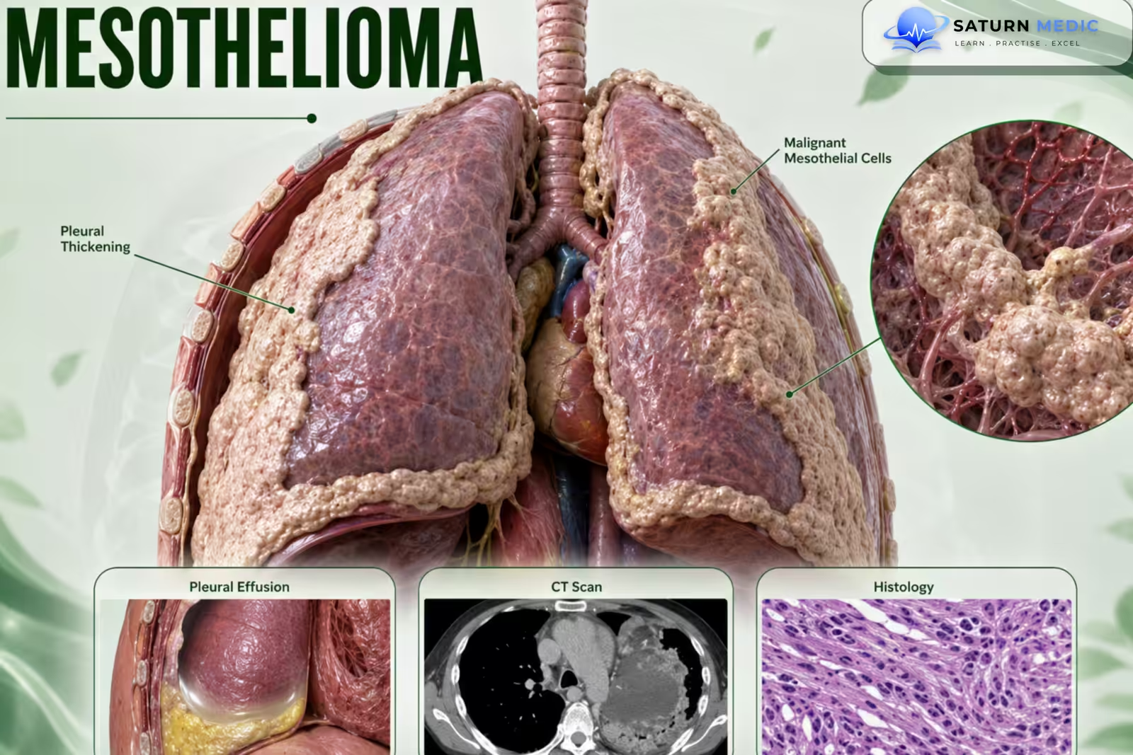
asbestos exposure. It has a long latency period and commonly presents with pleural
effusion, pleural thickening, dyspnea, and chest pain.
Etiology & Risk Factors
–Most important risk factor
• Asbestos exposure
-Other risk factors
• Exposure to other carcinogenic fibers (e.g., erionite)
• Genetic mutations (e.g., BAP1 mutation)
• Radiation exposure
Important: Smoking has not been shown to increase the incidence.
Pathophysiology
• Malignant transformation of mesothelial cells.
• Pleural mesothelioma is the most common type.
• Less common forms include:
• Peritoneal mesothelioma
• Pericardial mesothelioma
Clinical Presentation
-General features
• Long latency period (approximately 30–50 years after asbestos exposure)
• Fatigue
• Weight loss
• Fever
• Night sweats
-Pleural involvement
• Dyspnea
• Nonpleuritic chest pain (most common)
• Nonproductive cough
• Pleural effusion
• Dull percussion
• Reduced or absent breath sounds over the affected side
-Peritoneal involvement
• Features of ascites
-Other
• Paraneoplastic syndromes
History Taking
Assess for:
• Occupational asbestos exposure
• Duration since exposure
• Dyspnea
• Chest pain
• Cough
• Weight loss
• Fever
• Night sweats
• Symptoms of pleural effusion or ascites
Physical Examination
Assess for:
• Signs of pleural effusion
• Reduced breath sounds
• Dullness to percussion
• Respiratory status
• Constitutional features
• Evidence of ascites (if peritoneal disease)
Investigations
-Initial imaging
• Chest X-ray
• Contrast-enhanced CT chest (most sensitive imaging modality for diagnosis)
-CT findings
• Multiple nodular pleural lesions (pleural thickening)
• Pleural effusion
• Thickened interlobar fissures
• Pleural plaques or calcifications
• Reduced ipsilateral lung volume
• Mediastinal shift
• Signs of local invasion
-Pleural fluid analysis
Indicated in patients with pleural effusion.
Typical findings:
• Bloody exudative pleural fluid
• May contain malignant mesothelial cells
-Biopsy (Gold Standard)
Usually:
• Thoracoscopic biopsy
• (Laparoscopic biopsy for suspected peritoneal disease)
Purpose:
• Histopathological confirmation
• Differentiate mesothelioma from adenocarcinoma
• Determine subtype:
• Epithelioid
• Sarcomatoid
• Mixed
-Immunohistochemical markers include:
• Calretinin
• Cytokeratin 5 and 6
• Thrombomodulin
• Mesothelin
Diagnosis
Diagnosis is based on:
• History of asbestos exposure
• Clinical presentation
• Imaging findings
• Pleural fluid analysis
• Histopathological confirmation by biopsy (required)
Related Topics
Management of Mesothelioma
-Patients should be referred to:
• Oncology
• Pulmonology
• Experienced centers of excellence
-Systemic chemotherapy (Primary treatment)
First-line:
• Pemetrexed PLUS:
• Cisplatin, or
• Carboplatin
Selected patients:
• Bevacizumab may be added.
-Surgery
Selected patients with localized disease:
• Pleurectomy with decortication (preferred; lung-sparing)
• Pneumonectomy
-Radiation therapy
May be used as part of multimodal treatment.
-Palliative care
• Pleurodesis
• Indwelling pleural catheter for recurrent pleural effusions
• Palliative radiotherapy for pain or symptomatic obstruction
Complications of Mesothelioma
• Pleural effusion
• Local tumor invasion
• Metastasis (approximately 50% of cases)
• Pulmonary embolism
• Bronchopneumonia
• Cardiac tamponade
• Invasion of great vessels
• Cachexia
Prognosis of Mesothelioma
• Generally poor.
• Median survival in unresectable cases is approximately 1 year.
• About 50% of cases metastasize.
Key Points / Clinical Pearls of Mesothelioma
• Rare malignant tumor of mesothelial cells.
• Pleural mesothelioma is the most common type.
• Strongly associated with asbestos exposure.
• Latency period is 30–50 years.
• Presents with pleural effusion, dyspnea, and chest pain.
• Contrast-enhanced CT is the most sensitive initial imaging test.
• Histopathological confirmation by biopsy is essential.
• First-line treatment is pemetrexed plus cisplatin or carboplatin
- Syed HA, Wallen JM. National Center for Biotechnology Information (NIH). Malignant Mesothelioma, StatPearls.
- MedlinePlus, National Library of Medicine (NIH). Mesothelioma: Health Topic.
- Popat S, Baas P, Faivre-Finn C, et al; ESMO Guidelines Committee. Malignant Pleural Mesothelioma: ESMO Clinical Practice Guidelines for Diagnosis, Treatment and Follow-Up. Ann Oncol. 2022. PMID: 34861373.
- Myers DJ, Babiker HM. National Center for Biotechnology Information (NIH). Benign Mesothelioma, StatPearls.
- Bibby AC, Tsim S, Kanellakis N, et al. Malignant Pleural Mesothelioma: An Update on Investigation, Diagnosis and Treatment. Eur Respir Rev. 2016. PMID: 27903668.