

Clinical Subject Page
Gastric Cancer
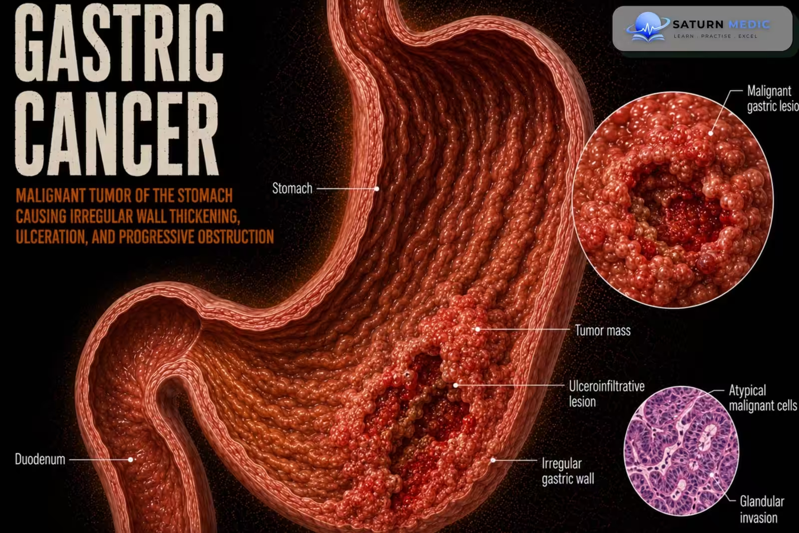
Gastric carcinoma is a malignant tumor of the stomach. Adenocarcinoma accounts for
approximately 95% of gastric cancers and is classified into intestinal and diffuse types
Also called
ICD-10
Specialty
Onset
Reviewed
-
OverviewOverview
-
Etiology & Risk FactorsEtiology & Risk Factors
-
PathophysiologyPathophysiology
-
Clinical PresentationClinical Presentation
-
History TakingHistory Taking
-
Physical ExaminationPhysical Examination
-
InvestigationsInvestigations
-
DiagnosisDiagnosis
-
ManagementManagement
-
ComplicationsComplications
-
PrognosisPrognosis
-
Key Points / Clinical PearlsKey Points / Clinical Pearls
Overview
Gastric cancer is the fifth most common cancer worldwide. Although its incidence has declined in
the United States, it remains common in several Asian countries. The major risk factor is
Helicobacter pylori infection. Early disease is often asymptomatic or presents with nonspecific
symptoms, resulting in delayed diagnosis and a poor overall prognosis.
Etiology & Risk Factors
Etiology
Gastric cancer is a malignant tumor arising from the gastric mucosa, with adenocarcinoma accounting for more than 90% of cases. It develops through a multistep process involving chronic inflammation, mucosal atrophy, intestinal metaplasia, dysplasia, and invasive carcinoma.
Risk Factors
Helicobacter pylori infection (most important risk factor)
Chronic atrophic gastritis
Intestinal metaplasia
Smoking
Family history of gastric cancer
Advanced age
Male sex
Diet high in smoked, salted, or preserved foods
Low fruit and vegetable intake
Previous partial gastrectomy
Pernicious anemia
Epstein–Barr virus infection
Hereditary diffuse gastric cancer (CDH1 mutation)
Familial adenomatous polyposis (FAP)
Lynch syndrome
Pathophysiology
1. Chronic exposure to carcinogenic or inflammatory factors leads to malignant transformation of
the gastric mucosa.
2. Adenocarcinoma (95%) develops and is classified as:
– Intestinal type
– Diffuse type
3. Tumor progressively invades the gastric wall.
4. Local invasion, lymphatic spread, and distant metastasis may occur.
Clinical Presentation
-
Early disease:
• Often asymptomatic
• Dyspepsia
• Epigastric pain
Advanced disease:
• Gastric outlet obstruction
• Gastrointestinal bleeding
• Ascites
• Features of metastatic disease
History Taking
-
Ask about:
• Dyspepsia
• Epigastric pain
• Duration and progression of symptoms
• Weight loss (if present)
• Symptoms of gastric outlet obstruction
• Gastrointestinal bleeding
• History of H. pylori infection
• Smoking history
• Dietary habits (high salt or nitrate intake)
• History of GERD
Physical Examination
Assess for:
• Epigastric tenderness
• Signs of gastrointestinal bleeding
• Ascites
• Clinical evidence of metastatic disease
• Nutritional status
Investigations
-
-
-
Initial Laboratory Tests
-
Complete blood count (CBC)
-
Liver function tests (LFTs)
-
Renal function tests
-
Electrolytes
-
Iron studies (if anemia is suspected)
Diagnostic Investigations
Upper Gastrointestinal Endoscopy (EGD) with Biopsy – Gold Standard
-
Gold standard investigation
-
Direct visualization of the lesion
-
Multiple biopsies for histopathological confirmation
Staging Investigations
-
Contrast-enhanced CT chest, abdomen, and pelvis
-
Endoscopic ultrasound (EUS) for T and N staging
-
PET-CT (selected patients)
-
Diagnostic laparoscopy for locally advanced disease to detect occult peritoneal metastases
Additional Investigations
-
HER2 testing
-
MSI/MMR testing
-
PD-L1 testing (advanced disease)
-
-
-
Diagnosis
-Diagnosis is confirmed by:
• Upper endoscopy demonstrating the lesion
• Histopathological examination of biopsy tissue
Tumor staging is based on:
• Imaging
• Diagnostic laparoscopy
Related Topics
Management
1. Early Gastric Cancer
-
Endoscopic mucosal resection (EMR)
-
Endoscopic submucosal dissection (ESD) (selected patients)
2. Localized Resectable Disease
-
Gastrectomy with lymph node dissection
-
Perioperative chemotherapy
-
Adjuvant chemotherapy when indicated
3. Advanced or Metastatic Disease
-
Systemic chemotherapy
-
Immunotherapy (selected patients)
-
HER2-targeted therapy (HER2-positive tumors)
-
Palliative radiotherapy or surgery when indicated
4. Supportive Care
-
Nutritional support
-
Pain management
-
Palliative care for advanced disease
5. Follow-Up
-
-
Regular clinical review
-
Surveillance imaging/endoscopy when indicated
-
Monitor for recurrence and nutritional deficiencies
-
Complications
• Gastric outlet obstruction
• Gastrointestinal bleeding
• Ascites
• Metastatic disease
Prognosis
- Prognosis depends largely on tumor stage at diagnosis.
- Overall survival is poor because many
patients are diagnosed only after symptoms develop and the disease has advanced
Key Points / Clinical Pearls
• Gastric carcinoma is the fifth most common cancer worldwide.
• Adenocarcinoma accounts for 95% of cases.
• The main risk factor is Helicobacter pylori infection.
• Other risk factors include GERD, smoking, and diets high in salts and nitrates.
• Early disease is often asymptomatic or causes nonspecific dyspepsia.
• Upper endoscopy with biopsy is the diagnostic test of choice.
• Imaging and diagnostic laparoscopy are used for staging.
• Early disease is treated with surgery and perioperative chemotherapy.
• Advanced disease is managed with systemic therapy, chemoradiation, targeted therapy, or
palliative care.
• Prognosis is generally poor due to late presentation
- National Center for Biotechnology Information (NIH). Gastric Cancer, StatPearls.
- Ajani JA, D'Amico TA, Bentrem DJ, et al. Gastric Cancer, Version 2.2025, NCCN Clinical Practice Guidelines in Oncology. J Natl Compr Canc Netw. 2025;23:169-191. PMID: 40341199.
- Dinis-Ribeiro M, Libânio D, Uchima H, et al. Management of Epithelial Precancerous Conditions and Early Neoplasia of the Stomach (MAPS III). Endoscopy. 2025. Endoscopy.
- National Cancer Institute (NIH). Gastric Cancer Treatment (PDQ), Health Professional Version.
- MedlinePlus, National Library of Medicine (NIH). Stomach Cancer: Health Topic.