

Clinical Subject Page
Achalasia
Achalasia is an esophageal motility disorder characterized by impaired relaxation of the lower
esophageal sphincter (LES) due to degeneration of inhibitory neurons within the esophageal wall.
Also called
ICD-10
Specialty
Onset
Reviewed
-
OverviewOverview
-
Etiology & Risk FactorsEtiology & Risk Factors
-
PathophysiologyPathophysiology
-
Clinical PresentationClinical Presentation
-
History TakingHistory Taking
-
Physical ExaminationPhysical Examination
-
InvestigationsInvestigations
-
DiagnosisDiagnosis
-
ManagementManagement
-
ComplicationsComplications
-
PrognosisPrognosis
-
Key Points / Clinical PearlsKey Points / Clinical Pearls
Overview
Achalasia causes failure of the LES to relax and impaired esophageal motility, resulting in
dysphagia, regurgitation, chest pain, and weight loss. High-resolution esophageal manometry is the
preferred test to confirm the diagnosis. Definitive treatment includes pneumatic dilation,
laparoscopic Heller myotomy, or peroral endoscopic myotomy (POEM).
Etiology & Risk Factors
Etiology
Most cases are idiopathic, caused by degeneration of inhibitory neurons in the myenteric (Auerbach) plexus, resulting in impaired relaxation of the lower esophageal sphincter (LES) and absent esophageal peristalsis.
Less common causes include:
Chagas disease (Trypanosoma cruzi infection)
Autoimmune-mediated neuronal injury
Genetic predisposition (rare)
Malignancy causing pseudoachalasia (e.g., gastroesophageal junction cancer)
Infiltrative disorders (rare)
Risk Factors
Age 25–60 years (can occur at any age)
Family history (rare)
Autoimmune diseases
Chagas disease (endemic regions of Latin America)
Esophagogastric junction malignancy (pseudoachalasia)
Rare genetic syndromes (e.g., Allgrove/Triple A syndrome)
Pathophysiology
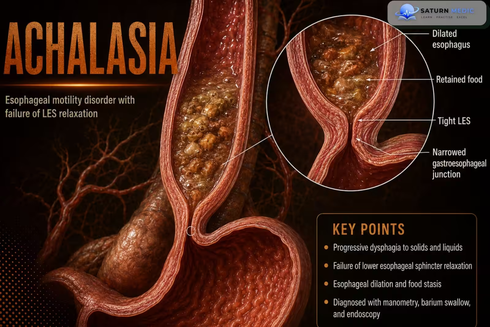
1. Degeneration of inhibitory neurons in the esophageal wall.
2. Impaired relaxation of the lower esophageal sphincter (LES).
3. Impaired esophageal emptying.
4. Progressive esophageal dilation with food retention.
5. Symptoms develop due to obstruction of food passage.
Clinical Presentation
Common symptoms:
• Dysphagia to both solids and liquids (most common)
• Regurgitation
• Retrosternal (chest) pain
• Weight loss
History Taking
Ask about:
• Dysphagia to solids and liquids
• Regurgitation of undigested food
• Retrosternal chest pain
• Weight loss
• Duration and progression of symptoms
• Previous treatments
• Symptoms suggesting malignancy (to assess for pseudoachalasia)
Physical Examination
- Physical examination is often normal.
- Assess nutritional status and evidence of weight loss.
Investigations
Initial Tests
Complete blood count (CBC)
Electrolytes and renal function tests
Nutritional assessment (if significant weight loss)
Diagnostic Investigations
Barium Swallow (Esophagram)
First-line investigation
Shows:
Bird’s beak narrowing at the gastroesophageal junction
Dilated esophagus
Delayed esophageal emptying
Upper Gastrointestinal Endoscopy (EGD)
Excludes mechanical obstruction or malignancy (pseudoachalasia)
May show retained food, saliva, and a dilated esophagus
High-Resolution Esophageal Manometry (Gold Standard)
Findings:
Impaired relaxation of the lower esophageal sphincter (LES)
Absent normal esophageal peristalsis
Classifies achalasia into Type I, II, and III
Additional Investigations
CT chest or abdomen if pseudoachalasia or malignancy is suspected
Endoscopic ultrasound (EUS) if gastroesophageal junction cancer is suspected
Diagnosis
Diagnosis is usually based on:
• Clinical features
• High-resolution esophageal manometry (gold-standard confirmatory test)
• Upper endoscopy to rule out pseudoachalasia
• Barium esophagram as an adjunct when indicated
Related Topics
Management
1. Definitive Therapy
Pneumatic Balloon Dilation (PD)
First-line option for many adults
Endoscopic balloon dilates the LES
May require repeat procedures
Laparoscopic Heller Myotomy (LHM)
Surgical division of the LES muscle
Usually combined with partial fundoplication to reduce postoperative reflux
Provides durable symptom relief
Peroral Endoscopic Myotomy (POEM)
Minimally invasive endoscopic myotomy
Particularly effective for type III (spastic) achalasia
Higher risk of postoperative GERD than Heller myotomy
2. Medical Therapy
Reserved for patients who are not candidates for definitive treatment.
Options include:
Nitrates (e.g., isosorbide dinitrate)
Calcium channel blockers (e.g., nifedipine)
Note: Symptom relief is usually temporary and less effective than procedural treatments.
3. Botulinum Toxin Injection
Endoscopic injection into the LES
Suitable for:
Elderly patients
Frail patients
Poor surgical candidates
Benefits are temporary and repeat injections are often required.
4. Nutritional Support
Eat slowly and chew food thoroughly
Drink water with meals
Eat smaller, frequent meals
Nutritional assessment if weight loss is significant
5. Follow-Up
Monitor symptom recurrence
Assess for GERD after myotomy or POEM
Repeat endoscopy if symptoms recur or complications are suspected
Long-standing achalasia may require surveillance for esophageal cancer in selected patients
Complications
• Progressive esophageal dilation
• Persistent dysphagia
• Weight loss
• Malnutrition
Prognosis
- Most patients experience symptomatic improvement with definitive treatment.
- Treatment choice
depends on the achalasia subtype, and recurrent symptoms may require reassessment.
Key Points / Clinical Pearls
• Achalasia is caused by degeneration of inhibitory neurons in the esophageal wall.
• It results in impaired LES relaxation.
• Dysphagia to both solids and liquids is the hallmark symptom.
• High-resolution esophageal manometry is the preferred diagnostic test.
• Upper endoscopy is required to exclude pseudoachalasia.
• Barium esophagram supports diagnosis and follow-up.
• Definitive treatments are POEM, laparoscopic Heller myotomy, and pneumatic dilation.
• Botulinum toxin is used when definitive therapy is not feasible.
• Nifedipine is considered only as a last-resort medical option.
- Puri R, Tian C. National Center for Biotechnology Information (NIH). Achalasia, StatPearls.
- Yadlapati R, Kahrilas PJ, Fox MR, et al. Esophageal Motility Disorders on High-Resolution Manometry: Chicago Classification Version 4.0. Neurogastroenterol Motil. 2021;33:e14058. PMID: 34490927.
- Vaezi MF, Pandolfino JE, Yadlapati RH, Greer KB, Kavitt RT. ACG Clinical Guidelines: Diagnosis and Management of Achalasia. Am J Gastroenterol. 2020;115:1393-1411. PMC9896940.
- MedlinePlus, National Library of Medicine (NIH). Achalasia: Medical Encyclopedia.
- National Center for Biotechnology Information (NIH). Esophageal Manometry, StatPearls.