

Clinical Subject Page
Celiac Disease
Esophageal carcinoma is a malignant tumor of the esophagus. The two major histological types are
esophageal adenocarcinoma and esophageal squamous cell carcinoma (SCC).
Also called
ICD-10
Specialty
Onset
Reviewed
-
OverviewOverview
-
Etiology & Risk FactorsEtiology & Risk Factors
-
PathophysiologyPathophysiology
-
Clinical PresentationClinical Presentation
-
History TakingHistory Taking
-
Physical ExaminationPhysical Examination
-
InvestigationsInvestigations
-
DiagnosisDiagnosis
-
ManagementManagement
-
ComplicationsComplications
-
PrognosisPrognosis
-
Key Points / Clinical PearlsKey Points / Clinical Pearls
Overview
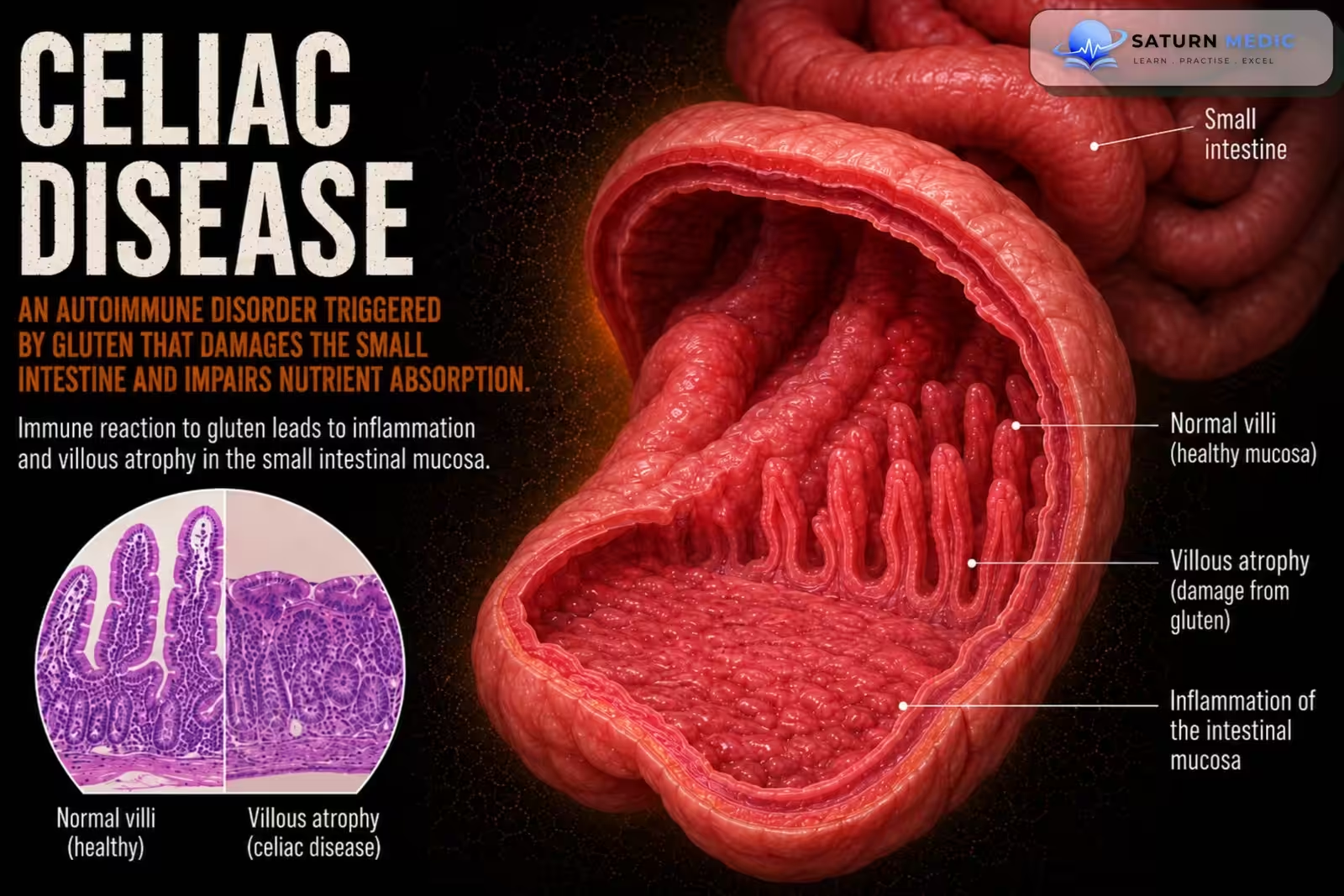
- The immune system attacks the small intestine after gluten exposure.
- This causes villous atrophy (flattening of the intestinal villi), leading to poor nutrient absorption (malabsorption).
Etiology & Risk Factors
Celiac disease is caused by a maladaptive immune response to gluten (a protein found in grains such as wheat) in genetically predisposed individuals.
The underlying mechanism involves:
- Gluten intolerance triggering an autoimmune reaction
- Production of autoantibodies against tissue transglutaminase (tTG) in the proximal small intestine
- Association with HLA variants that predispose to excessive immune responses
Risk factors
Factors associated with an increased risk of celiac disease include:
- Genetic predisposition, particularly HLA-DQ2 and HLA-DQ8
- Female sex (♀ > ♂)
- Northern European ancestry
- Presence of other autoimmune diseases, as these share HLA-associated immune susceptibility
Additional epidemiologic features:
- Can occur at any age
- Peak incidence:
- 8–12 months of age (typically 2–3 months after first gluten exposure)
- Third to fourth decade of life
Pathophysiology
Gluten ingestion (gliadin) → tTG modifies gliadin → Immune (T-cell) activation in genetically susceptible individuals → Chronic inflammation of the small intestine → Villous atrophy + crypt hyperplasia + brush border loss → Malabsorption → Nutrient deficiencies and gastrointestinal symptoms
Clinical Presentation
- Gastrointestinal Symptoms
- Chronic or recurrent diarrhea
- Steatorrhea (fatty stools)
- Abdominal bloating, flatulence, and pain
- Nausea/vomiting
- Poor appetite
- Constipation (less common)
Extraintestinal Symptoms
- Fatigue
- Weight loss
- Iron deficiency anemia and other vitamin deficiencies
- Osteoporosis or hypocalcemia
- Dermatitis herpetiformis (itchy blistering rash)
- Peripheral neuropathy, headache, ataxia, depression, irritability
Children
- Failure to thrive
- Growth failure
- Delayed puberty
History Taking
Ask about:
- Do you have chronic diarrhea or loose stools?
- Are your stools greasy, bulky, or difficult to flush?
- Do you have abdominal pain, bloating, or excessive gas?
- Have you had unexplained weight loss?
- Do you often feel tired or weak?
- Have you been told you have iron deficiency anemia or other vitamin deficiencies?
- Do your symptoms worsen after eating foods containing wheat, barley, or rye?
- Do you have an itchy, blistering skin rash?
- Is there a family history of celiac disease?
- Do you or your family have other autoimmune diseases (e.g., type 1 diabetes or thyroid disease)?
Physical Examination
- Weight loss or low BMI
- Pallor (suggesting anemia)
- Signs of dehydration (if severe diarrhea)
- Abdominal distension/bloating
- Abdominal tenderness (may be mild)
- Dermatitis herpetiformis (itchy, blistering rash)
- Signs of malnutrition (muscle wasting)
- Growth failure or delayed puberty (in children)
- Signs of vitamin deficiencies (e.g., glossitis, osteoporosis)
Investigations
Laboratory Tests
- tTG-IgA (tissue transglutaminase IgA) – first-line screening test
- Total serum IgA – check for IgA deficiency
- If IgA deficient:
- tTG-IgG
- Deamidated gliadin peptide (DGP-IgG)
- Endomysial antibody (EMA) – confirmatory serologic test
- Screen for nutrient deficiencies:
- CBC, iron/ferritin
- Folate, vitamin B12
- Calcium, vitamin D, zinc
Confirmatory Test
- Upper GI endoscopy (EGD) with multiple duodenal biopsies
- Shows villous atrophy, crypt hyperplasia, and increased intraepithelial lymphocytes
Additional Tests (if needed)
- HLA-DQ2/HLA-DQ8 genetic testing
- Gluten challenge (if already on a gluten-free diet before testing)
Diagnosis
- Diagnosis is based on:
- Clinical suspicion (symptoms or risk factors)
- Positive serology:
- tTG-IgA (first-line)
- Total IgA
- ± EMA or DGP-IgG (if IgA deficient)
- Confirmation: Upper GI endoscopy (EGD) with duodenal biopsy
- Shows villous atrophy, crypt hyperplasia, and increased intraepithelial lymphocytes
Related Topics
Management
Main Treatment :
- Strict lifelong gluten-free diet(cornerston)
- Avoid wheat, rye, barley, and spelt
- Eat naturally gluten-free foods (e.g., rice, corn, potatoes, soybeans, millet)
- Supportive Management
- Replace iron and vitamin/mineral deficiencies (e.g., iron, vitamin B12, folate, vitamin D, calcium)
- Avoid milk products temporarily if secondary lactose intolerance is present
- Refer to a dietitian for nutrition counseling
- Consider osteoporosis screening in adults
- Regular follow-up to assess symptom improvement and dietary adherence
Complications
- Malnutrition and nutrient deficiencies
- Secondary lactose intolerance (lactase deficiency)
- Osteoporosis/osteopenia (from malabsorption)
- Iron deficiency anemia
- Refractory celiac disease (persistent symptoms despite a strict gluten-free diet)
- Ulcerative jejunitis (in severe refractory disease)
- Increased risk of malignancy, especially:
- Enteropathy-associated T-cell lymphoma (EATL)
- Small bowel adenocarcinoma
Prognosis
- Excellent prognosis with a strict lifelong gluten-free diet
- Symptoms usually improve quickly, and intestinal healing occurs with continued dietary adherence.
- A gluten-free diet reduces the risk of complications, including intestinal lymphoma.
- Poor adherence to the diet increases the risk of persistent symptoms, malabsorption, and long-term complications.
Key Points / Clinical Pearls
- Autoimmune disease triggered by gluten (wheat, rye, barley, spelt).
- Causes damage to the small-intestinal villi → malabsorption.
- Common symptoms: chronic diarrhea, bloating, weight loss, fatigue, and iron deficiency anemia.
- First-line test: tTG-IgA + total IgA.
- Confirm diagnosis: Upper GI endoscopy with duodenal biopsy.
- Do not start a gluten-free diet before diagnostic testing if possible.
- Treatment: Strict lifelong gluten-free diet.
- Correct vitamin and mineral deficiencies.
- Untreated disease can lead to osteoporosis, malnutrition, infertility, and intestinal lymphoma.
- Prognosis is excellent with good adherence to a gluten-free diet.
- National Center for Biotechnology Information (NIH). Celiac Disease, StatPearls.
- Rubio-Tapia A, Hill ID, Semrad C, et al. American College of Gastroenterology Guidelines: Diagnosis and Management of Celiac Disease. Am J Gastroenterol. 2023;118:59-76. ACG Guideline.
- MedlinePlus, National Library of Medicine (NIH). Celiac Disease: Health Topic.
- National Center for Biotechnology Information (NIH). Gluten-Associated Medical Problems, StatPearls.
- National Center for Biotechnology Information (NIH). Dermatitis Herpetiformis, StatPearls.