

Clinical Subject Page
Splenomegaly
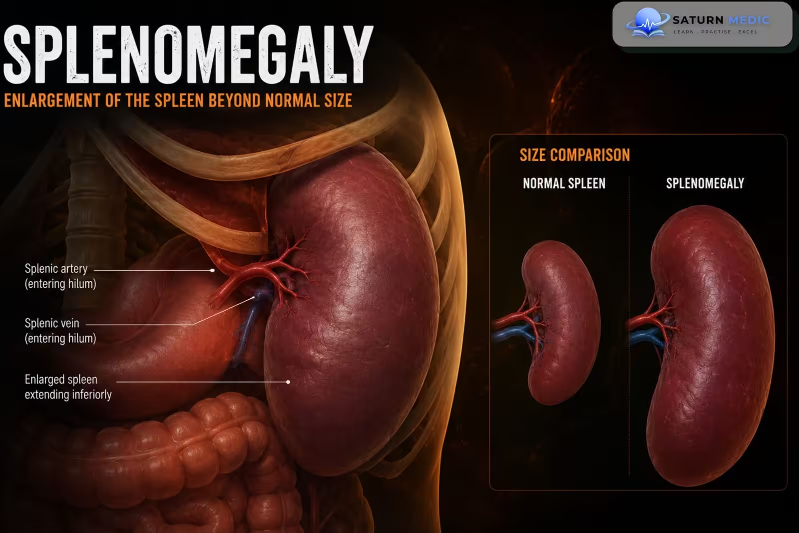
Splenomegaly is an abnormal enlargement of the spleen. It is not a disease by itself but usually indicates an underlying infection, liver disease, hematological disorder, malignancy, or systemic inflammatory condition.
Also called
ICD-10
Specialty
Onset
Reviewed
-
OverviewOverview
-
Etiology & Risk FactorsEtiology & Risk Factors
-
PathophysiologyPathophysiology
-
Clinical PresentationClinical Presentation
-
History TakingHistory Taking
-
Physical ExaminationPhysical Examination
-
InvestigationsInvestigations
-
DiagnosisDiagnosis
-
ManagementManagement
-
ComplicationsComplications
-
PrognosisPrognosis
-
Key Points / Clinical PearlsKey Points / Clinical Pearls
Overview
Splenomegaly develops when the spleen enlarges because of increased immune activity, blood-cell destruction, venous congestion, abnormal cell infiltration, or increased blood-cell production. Patients may be asymptomatic or present with left upper-quadrant discomfort, early satiety, anemia, recurrent infections, or easy bleeding. Ultrasound is commonly used to confirm enlargement, while further investigations aim to identify the underlying cause.
Etiology & Risk Factors
Aetiology and Risk Factors
-Liver and Vascular Causes
Liver cirrhosis
Portal hypertension
Portal vein thrombosis
Splenic vein thrombosis
Congestive heart failure
Budd–Chiari syndrome
-Infectious Causes
Infectious mononucleosis
Viral hepatitis
HIV infection
Malaria
Tuberculosis
Brucellosis
Infective endocarditis
Visceral leishmaniasis
-Hematological Causes
Hemolytic anemia
Sickle cell disease
Thalassemia
Hereditary spherocytosis
Autoimmune hemolytic anemia
Myelofibrosis
Polycythemia vera
-Malignant Causes
Leukemia
Lymphoma
Myeloproliferative neoplasms
Metastatic malignancy
-Autoimmune and Inflammatory Causes
Rheumatoid arthritis
Felty syndrome
Systemic lupus erythematosus
Sarcoidosis
Systemic inflammatory disorders
Pathophysiology
Underlying disease → increased splenic workload, venous congestion, abnormal cellular infiltration, or excessive blood-cell sequestration → enlargement of the spleen → increased trapping and destruction of blood cells → possible hypersplenism and cytopenias.
Clinical Presentation
Common symptoms include:
- Left upper-quadrant discomfort or pain
- Feeling of fullness beneath the left ribs
- Early satiety due to pressure on the stomach
- Abdominal fullness
- Fatigue
- Weight loss
- Fever or night sweats
- Easy bruising or bleeding
- Recurrent infections
History Taking
Ask about:
- Left upper-quadrant discomfort or fullness
- Early satiety
- Fever or recent infection
- Weight loss or night sweats
- Fatigue or pallor
- Easy bruising or bleeding
- Recurrent infections
- Jaundice or dark urine
- Abdominal swelling
- Alcohol use
- Known liver disease
- Recent travel to malaria-endemic regions
- Hematological disease or malignancy
- Autoimmune disease
- Recent abdominal trauma
Physical Examination
General Examination
Look for:
Pallor
Jaundice
Fever
Weight loss
Lymphadenopathy
Bruising or petechiae
Signs of chronic liver disease
Abdominal Examination
Assess for:
Palpable spleen below the left costal margin
Splenic tenderness
Hepatomegaly
Ascites
Abdominal distension
Signs of portal hypertension
An enlarged spleen typically moves downward with inspiration and may have a palpable notch along its medial border.
Other Examination
Look for:
Cardiac murmurs suggesting infective endocarditis
Joint inflammation
Skin rashes
Peripheral edema
Signs of hematological malignancy
Investigations
Initial Laboratory Tests
Complete blood count
Peripheral blood smear
Reticulocyte count
Liver function tests
Renal function and electrolytes
C-reactive protein and ESR
Coagulation profile
Lactate dehydrogenase
Bilirubin
Haptoglobin
Viral serology when indicated
Blood cultures if infection is suspected
Additional Tests Based on Suspected Cause
Malaria blood film
Monospot test or EBV serology
HIV testing
Hepatitis serology
Autoimmune screening
Hemoglobin electrophoresis
JAK2 mutation testing
Bone marrow examination
Flow cytometry
Imaging
Abdominal Ultrasound
Usually the first imaging test because it can:
Confirm splenic enlargement
Measure spleen size
Assess liver disease
Detect portal hypertension
Identify focal splenic lesions
CT Abdomen
Used when:
Malignancy is suspected
Splenic lesions are present
Complications are suspected
More detailed abdominal assessment is required
Doppler Ultrasound
May assess:
Portal vein thrombosis
Splenic vein thrombosis
Portal blood flow
Diagnosis
Diagnosis requires:
- Confirmation of splenic enlargement by examination or imaging
- Identification of the underlying cause
Splenomegaly should not be considered the final diagnosis until liver disease, infection, hematological disease, malignancy, and autoimmune conditions have been assessed.
Related Topics
Management
General Management
Treat the underlying cause
Avoid contact sports and abdominal trauma
Provide analgesia when needed
Monitor blood counts
Manage anemia, thrombocytopenia, or infection
Refer to hematology or hepatology when indicated
Cause-Specific Management
Antibiotics for bacterial infection
Antiparasitic treatment for malaria or leishmaniasis
Antiviral or supportive care for viral infections
Treatment of portal hypertension
Management of cirrhosis
Chemotherapy or targeted therapy for hematological malignancy
Immunosuppressive therapy for selected autoimmune disorders
Treatment of hemolytic anemia
Splenectomy
Splenectomy is rarely used only to treat splenomegaly itself. It may be considered for:
Severe hypersplenism
Symptomatic massive splenomegaly
Recurrent splenic complications
Selected hematological disorders
Suspected or confirmed splenic malignancy
Splenic rupture
Complications
- Hypersplenism
- Anemia
- Leukopenia
- Thrombocytopenia
- Recurrent infection
- Easy bleeding
- Splenic infarction
- Splenic rupture
- Abdominal discomfort
- Early satiety and poor nutrition
Prognosis
The prognosis depends on the underlying cause. Mild enlargement related to a temporary infection may resolve completely, while splenomegaly caused by cirrhosis, hematological malignancy, or chronic inflammatory disease may persist and require long-term management.
Key Points / Clinical Pearls
- Splenomegaly means abnormal enlargement of the spleen.
- It is usually secondary to another disease.
- Common causes include liver disease, infection, hemolytic anemia, and hematological malignancy.
- Patients may have left upper-quadrant discomfort or early satiety.
- CBC, blood smear, liver tests, and abdominal ultrasound are important initial investigations.
- Management focuses on treating the underlying cause.
- Patients should avoid abdominal trauma because an enlarged spleen is more vulnerable to rupture.
- Chapman J, Goyal A, Azevedo AM. National Center for Biotechnology Information (NIH). Splenomegaly, StatPearls.
- MedlinePlus, National Library of Medicine (NIH). Splenomegaly: Medical Encyclopedia.
- National Center for Biotechnology Information (NIH). Physiology, Spleen, StatPearls.
- National Center for Biotechnology Information (NIH). Asplenia, StatPearls.
- National Center for Biotechnology Information (NIH). Spleen Imaging, StatPearls.