

Clinical Subject Page
Anal Cancer
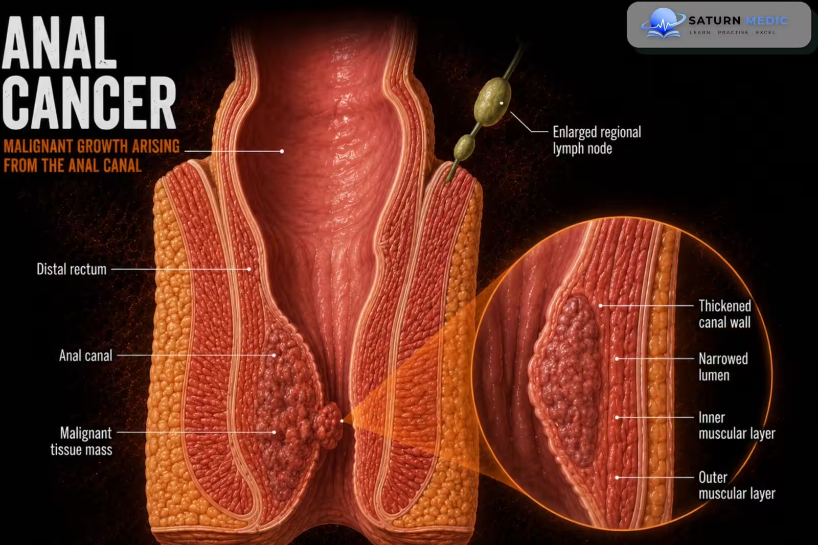
Anal cancer is a malignant tumor arising from the anal canal or anal margin, with
squamous cell carcinoma (SCC) accounting for the vast majority of cases
Also called
ICD-10
Specialty
Onset
Reviewed
-
OverviewOverview
-
Etiology & Risk FactorsEtiology & Risk Factors
-
PathophysiologyPathophysiology
-
Clinical PresentationClinical Presentation
-
History TakingHistory Taking
-
Physical ExaminationPhysical Examination
-
InvestigationsInvestigations
-
DiagnosisDiagnosis
-
ManagementManagement
-
ComplicationsComplications
-
PrognosisPrognosis
-
Key Points / Clinical PearlsKey Points / Clinical Pearls
Overview
Anal cancer is an uncommon gastrointestinal malignancy strongly associated with persistent human papillomavirus (HPV) infection, particularly HPV-16. Most patients present with rectal bleeding, anal pain, or a palpable mass. Diagnosis is confirmed by biopsy, and the standard treatment for most anal canal cancers is combined chemoradiotherapy, which preserves anal sphincter function
Etiology & Risk Factors
Etiology
Most anal cancers develop from persistent high-risk HPV infection, leading to anal
intraepithelial neoplasia (AIN) and progression to invasive squamous cell carcinoma.
Risk Factors
– Persistent HPV infection (especially HPV-16)
– HIV infection
– Immunosuppression
– Receptive anal intercourse
– Multiple sexual partners
– Smoking
– History of cervical, vulvar, or vaginal dysplasia/cancer
– Men who have sex with men (MSM)
– Chronic immunosuppressive therapy
– Increasing age
Pathophysiology
Persistent infection with high-risk human papillomavirus, especially HPV-16 and HPV-18, causes integration of viral DNA into anal squamous epithelial cells → viral oncoproteins E6 and E7 inactivate the tumor suppressor proteins p53 and retinoblastoma protein → abnormal cell-cycle progression and accumulation of genetic mutations → development of anal intraepithelial neoplasia → progression from low-grade to high-grade dysplasia → invasion through the basement membrane → formation of anal squamous cell carcinoma → local spread into the sphincters, rectum, perianal tissues, and regional lymph nodes.
Clinical Presentation
Common Symptoms
– Bright red rectal bleeding
– Anal pain
– Anal mass
– Change in bowel habits
– Pruritus ani
– Mucous discharge
– Tenesmus
– Pain during defecation
Advanced Disease
– Enlarging anal mass
– Inguinal lymphadenopathy
– Weight loss
– Persistent ulcer
– Fecal incontinence
History Taking
Ask about: Rectal bleeding, anal pain, anal lump, change in bowel habits, weight loss, mucous discharge, HIV status, HPV history, smoking, previous HPV-related cancers, immunosuppression
Physical Examination
General examination:
weight loss, performance status.Perianal examination: ulcer, anal mass, bleeding, induration, perianal skin lesions.
Digital rectal examination:
tumor size, fixation, sphincter involvement, tenderness.
Inguinal examination: palpate inguinal lymph nodes.
Anoscopy:
direct visualization, determine tumor location, obtain biopsy.
Investigations
Laboratory Tests
CBC, renal function, liver function tests, HIV testing (when appropriate).
Tissue Diagnosis Biopsy (Gold Standard):
confirms malignancy and histological subtype.
Imaging for Staging
Pelvic MRI (Preferred Local Staging): tumor size, sphincter involvement, local invasion.
CT Chest, Abdomen & Pelvis: nodal and distant metastases.
PET-CT: nodal disease, occult metastases, treatment planning.
Endoscopic Assessment
Anoscopy and proctoscopy.
Important Note
Biopsy is the gold standard for diagnosis. Pelvic MRI is preferred for local staging, while
CT and PET-CT are used to assess regional and distant spread.
Diagnosis
Based on history, physical examination, digital rectal examination, anoscopy with
biopsy, MRI for local staging, and CT and/or PET-CT for staging.
Related Topics
Management
1. Localized Anal Canal Cancer (First-Line)
Combined chemoradiotherapy: 5-Fluorouracil (5-FU) or Capecitabine, Mitomycin,
External beam radiotherapy.
2. Persistent or Recurrent Disease
Salvage abdominoperineal resection (APR).
3. Metastatic Disease
Systemic chemotherapy, immunotherapy (selected patients), palliative radiotherapy when indicated.
4. Follow-Up
Clinical examination, digital rectal examination, anoscopy, imaging when indicated.
Important Note
Combined chemoradiotherapy is the standard treatment for most anal canal cancers
and usually avoids the need for permanent colostomy. Surgery is mainly reserved for
persistent or recurrent disease
Complications
– Local recurrence
– Inguinal lymph node metastasis
– Distant metastasis
– Anal stenosis
– Fecal incontinence
– Sexual dysfunction
– Chronic radiation toxicity
Prognosis
– Excellent for early-stage disease
– Most patients achieve complete remission with chemoradiotherapy
– Prognosis depends on tumor size, nodal involvement, and metastatic disease
– Early diagnosis significantly improves survival
Key Points / Clinical Pearls
– Most anal cancers are squamous cell carcinomas.
– Persistent HPV infection is the major risk factor.
– Rectal bleeding should not automatically be attributed to hemorrhoids.
– Biopsy is the gold standard for diagnosis.
– Pelvic MRI is preferred for local staging.
– Combined chemoradiotherapy is first-line treatment for most anal canal cancers.
– Surgery (APR) is mainly reserved for persistent or recurrent disease.
– Regular follow-up is essential to detect recurrence.
- National Center for Biotechnology Information (NIH). Radiation Therapy for Anal Cancer, StatPearls.
- National Comprehensive Cancer Network (NCCN). NCCN Clinical Practice Guidelines in Oncology: Anal Carcinoma, Version 2.2026.
- National Cancer Institute (NIH). Anal Cancer Treatment (PDQ), Health Professional Version.
- MedlinePlus, National Library of Medicine (NIH). Anal Cancer: Health Topic.
- Leeds IL, Fang SH. Anal Cancer and Intraepithelial Neoplasia Screening: A Review. World J Gastrointest Surg. 2016;8:41-51.