

Clinical Subject Page
Bronchiectasis
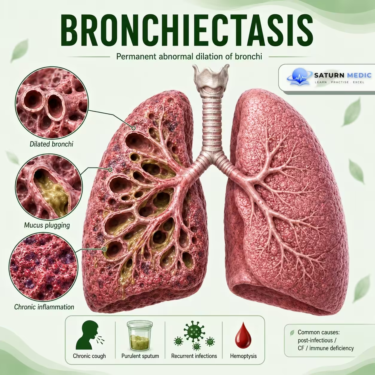
Bronchiectasis is a chronic lung disease characterized by permanent abnormal widening of the bronchi, leading to impaired mucus clearance, recurrent infections, and chronic airway inflammation.
Also called
ICD-10
Specialty
Onset
Reviewed
-
OverviewOverview
-
Etiology & Risk FactorsEtiology & Risk Factors
-
PathophysiologyPathophysiology
-
Clinical PresentationClinical Presentation
-
History TakingHistory Taking
-
Physical ExaminationPhysical Examination
-
InvestigationsInvestigations
-
DiagnosisDiagnosis
-
ManagementManagement
-
ComplicationsComplications
-
PrognosisPrognosis
-
Key Points / Clinical PearlsKey Points / Clinical Pearls
Overview
Damaged and widened bronchi cannot clear mucus effectively. Mucus accumulates, promoting recurrent infection and further airway damage.
Etiology & Risk Factors
Common Causes
- Previous severe lung infection
- Cystic fibrosis
- Primary ciliary dyskinesia
- Immune deficiency
- Allergic bronchopulmonary aspergillosis (ABPA)
- Airway obstruction
- Autoimmune disease
- Aspiration
Important Note
In many patients, no specific cause is identified (idiopathic bronchiectasis).
-Risk Factors
- Recurrent respiratory infections
- Previous tuberculosis
- Immunodeficiency
- Chronic aspiration
- Severe childhood lung infections
- Underlying genetic disorders
Pathophysiology
Initial airway injury
⬇
Impaired mucus clearance
⬇
Mucus accumulation
⬇
Bacterial infection
⬇
Chronic inflammation
⬇
Airway wall destruction
⬇
Permanent bronchial dilatation
⬇
Further mucus retention and recurrent infection
Key Concept
This forms a vicious cycle of infection, inflammation, and airway damage.
Clinical Presentation
Symptoms
Chronic productive cough
Daily sputum production
Recurrent chest infections
Dyspnea
Wheezing
Hemoptysis
Fatigue
Sputum
May be:
Large in volume
Purulent
Foul-smelling during infection
Signs
Coarse crackles
Wheezing
Digital clubbing in advanced disease
Reduced oxygen saturation in severe disease
History Taking
Ask about:
- Chronic cough?
- Daily sputum?
- Sputum amount and color?
- Recurrent chest infections?
- Hemoptysis?
- Dyspnea or wheezing?
- Previous tuberculosis or severe pneumonia?
- Frequent childhood infections?
- Recurrent sinus or ear infections?
- Aspiration symptoms?
Physical Examination
-General Examination
Look for:
- Respiratory distress
- Cyanosis
- Digital clubbing
- Reduced oxygen saturation
-Chest Examination
- Coarse inspiratory crackles
- Wheezing
- Rhonchi
- Reduced breath sounds in advanced disease
Investigations
-High-Resolution CT (HRCT) Chest — Key Test
May show:
Dilated bronchi
Lack of normal bronchial tapering
Bronchi larger than accompanying pulmonary arteries
Bronchial wall thickening
-Sputum Tests
Sputum culture
Antibiotic sensitivity
Mycobacterial testing when indicated
-Pulmonary Function Tests
May show:
Obstructive pattern
Mixed pattern
Sometimes normal results
-Blood Tests
CBC
Immunoglobulin levels
Tests for ABPA
Autoimmune testing when indicated
-Additional Tests
Depending on the suspected cause:
Cystic fibrosis testing
Ciliary function testing
Bronchoscopy
Diagnosis
Bronchiectasis · Stepwise Diagnostic Approach — ERS 2017
Related Topics
Management
Bronchiectasis · Treatment — ERS Guidelines 2017
| Indication | First-line antibiotic | Alternative / P. aeruginosa | Duration & notes |
|---|---|---|---|
| Mild exacerbation Oral |
Amoxicillin 500 mg–1g TDS Amoxicillin-clavulanate 625 mg TDS |
Doxycycline 100 mg BD (if penicillin allergy) Azithromycin 500 mg OD |
14 days. Guided by previous sputum cultures. Start empirically while awaiting cultures. |
| Moderate–severe exacerbation IV |
Co-amoxiclav IV Ceftriaxone IV 2g OD |
Piperacillin-tazobactam IV Meropenem IV |
14 days IV; step down to oral when clinical improvement. Hospital admission for IV treatment. |
| P. aeruginosa colonisation — exacerbation Targeted |
Ciprofloxacin 500–750 mg BD oral (if susceptible) |
Piperacillin-tazobactam IV Ceftazidime IV Meropenem IV |
14 days. Dual IV therapy sometimes used for severe P. aeruginosa. Always guided by sensitivity. |
| Long-term suppressive antibiotics Chronic |
Azithromycin 250–500 mg 3x/week (most evidence) Erythromycin 250 mg BD |
Inhaled colistin Inhaled tobramycin Inhaled aztreonam |
Indicated if ≥3 exacerbations/year. Azithromycin reduces exacerbations ~30%. Monitor QTc, hearing (macrolides). Screen for NTM before starting. |
| New P. aeruginosa isolation — eradication Eradication |
Ciprofloxacin 500–750 mg BD × 2–3 weeks | Inhaled colistin + oral ciprofloxacin | Attempt eradication on first isolation of P. aeruginosa. Repeat sputum at 3 months to confirm clearance. Success ~30–50%. |
Complications
- Recurrent pneumonia
- Frequent exacerbations
- Massive hemoptysis
- Chronic respiratory failure
- Pulmonary hypertension
- Cor pulmonale
- Progressive loss of lung function
- Reduced quality of life
Prognosis
- The course is highly variable.
- Many patients remain stable with appropriate treatment.
- Prognosis is worse with:
- Frequent exacerbations
- Chronic bacterial infection
- Extensive lung involvement
- Poor lung function
- Respiratory failure
- Early diagnosis and effective airway clearance can improve long-term outcomes.
Key Points / Clinical Pearls
- Bronchiectasis is permanent abnormal dilatation of the bronchi.
- Chronic productive cough is the classic symptom.
- Recurrent chest infections are common.
- HRCT is the key test for diagnosis.
- Sputum culture helps guide antibiotic treatment.
- Airway clearance is a cornerstone of management.
- Hemoptysis may occur and can be severe.
- Bird K, Memon J. National Center for Biotechnology Information (NIH). Bronchiectasis, StatPearls.
- Chalmers JD, Polverino E, Aliberti S, et al; ERS Task Force. European Respiratory Society Clinical Practice Guideline for the Management of Adult Bronchiectasis. Eur Respir J. 2025;66:2501126. doi: 10.1183/13993003.01126-2025.
- MedlinePlus, National Library of Medicine (NIH). Bronchiectasis: Medical Encyclopedia.
- Lonni S, Chalmers JD, Goeminne PC, et al. Etiology of Non-Cystic Fibrosis Bronchiectasis in Adults and Its Correlation to Disease Severity. Ann Am Thorac Soc. 2015. PMID: 26421502.
- Hill AT, Sullivan AL, Chalmers JD, et al. British Thoracic Society Guideline for Bronchiectasis in Adults. Thorax. 2019. PMID: 30442718.