Saturn Medic

Clinical Subject Page

Chronic obstructive pulmonary disease (COPD)

Chronic obstructive pulmonary disease (COPD) is a chronic lung disease characterized by persistent respiratory symptoms and airflow obstruction caused by abnormalities of the airways and/or alveoli.

Also called

Chronic obstructive lung disease (COLD)

ICD-10

J44.9

Specialty

Pulmonology

Onset

Chronic

Reviewed

July 2026

On This Page

Overview

COPD develops gradually, usually after long-term exposure to cigarette smoke or other harmful particles and gases.

It mainly includes features of:

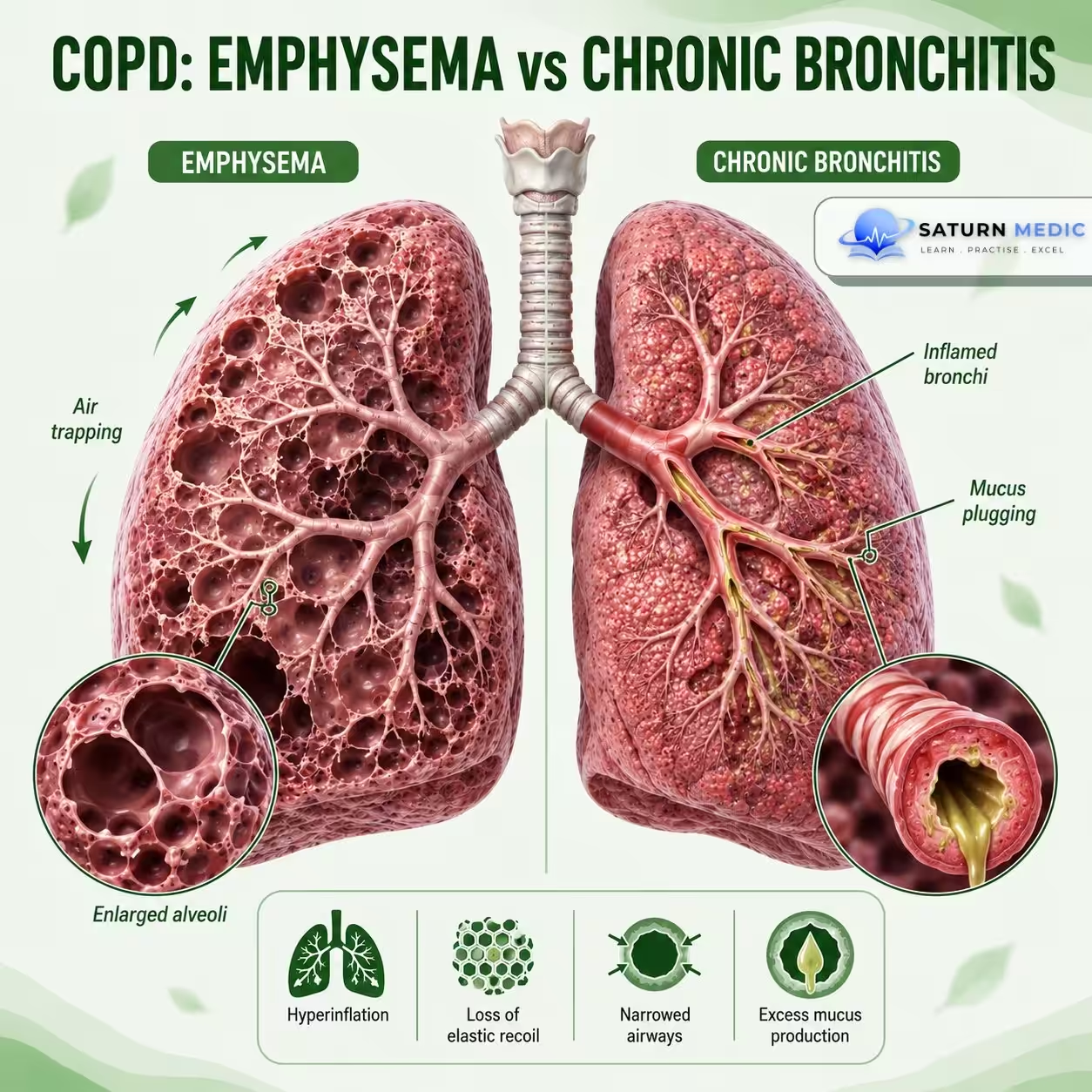
  • Chronic bronchitis → airway inflammation and mucus production
  • Emphysema → destruction of alveoli and loss of elastic recoil

The airflow obstruction is persistent and usually progressive.

Etiology & Risk Factors

Main Cause

Cigarette Smoking

The most important risk factor for COPD.

Other Causes

  • Biomass fuel exposure
  • Occupational dusts and chemicals
  • Air pollution
  • Recurrent respiratory infections
  • Abnormal lung development

Genetic Cause

Alpha-1 Antitrypsin Deficiency

An important inherited cause, especially in:

  • Younger patients
  • Patients with little or no smoking history
  • Patients with early-onset emphysema

Risk Factors

  • Smoking
  • Increasing age
  • Occupational exposure
  • Indoor air pollution
  • Outdoor air pollution
  • Poor lung growth
  • Family history of COPD

Pathophysiology

Long-term exposure to harmful particles or gases

Chronic airway inflammation

Small airway narrowing + Mucus hypersecretion

Alveolar wall destruction

Loss of elastic recoil

Air trapping and hyperinflation

Persistent expiratory airflow limitation

Dyspnea + Cough + Sputum production

Key Concept

Unlike asthma, airflow obstruction in COPD is persistent and not fully reversible.

Clinical Presentation

  1. Symptoms

    • Progressive dyspnea
    • Chronic cough
    • Sputum production
    • Wheezing
    • Chest tightness
    • Reduced exercise tolerance
    • Fatigue

    Typical Pattern

    Symptoms usually:

    • Develop gradually
    • Progress over years
    • Worsen with exertion
    • Become worse during exacerbations

    Signs

    • Prolonged expiration
    • Wheezing
    • Reduced breath sounds
    • Hyperinflated chest
    • Accessory muscle use
    • Pursed-lip breathing
    • Cyanosis in advanced disease
    • Peripheral edema in cor pulmonale

History Taking

  • Ask about:

    • Shortness of breath?
    • Chronic cough?
    • Daily sputum?
    • Wheezing?
    • When did symptoms start?
    • Are symptoms progressive?
    • Exercise limitation?
    • Smoking history?
    • Pack-years?
    • Biomass fuel exposure?
    • Occupational exposure?
    • Previous exacerbations?
    • Previous hospital admissions?
    • Current inhalers?
    • Inhaler technique?
    • Treatment adherence?

Physical Examination

  • Look for:

    • Respiratory distress
    • Tachypnea
    • Cyanosis
    • Accessory muscle use
    • Pursed-lip breathing
    • Reduced oxygen saturation
    • Low body weight in advanced disease

    Chest Examination

    • Barrel-shaped chest
    • Reduced chest expansion
    • Hyperresonance
    • Reduced breath sounds
    • Prolonged expiration
    • Wheezing

    Advanced Disease

    Look for:

    • Raised JVP
    • Peripheral edema
    • Signs of pulmonary hypertension
    • Signs of cor pulmonale

Investigations

  • Spirometry — Key Test

    Shows:

    • Persistent airflow obstruction
    • Reduced FEV₁/FVC ratio

    Post-bronchodilator spirometry is required to confirm persistent airflow obstruction.

    Pulse Oximetry

    Assesses:

    • Oxygen saturation

    Arterial Blood Gas

    Consider in:

    • Severe COPD
    • Severe exacerbations
    • Suspected respiratory failure

    Chest X-ray

    May show:

    • Hyperinflation
    • Flattened diaphragm
    • Increased lung lucency

    CT Chest

    Consider when:

    • Diagnosis is uncertain
    • Emphysema needs assessment
    • Lung cancer is suspected
    • Surgery or other interventions are being considered

    Blood Tests

    • CBC
    • Alpha-1 antitrypsin testing when indicated

Diagnosis

COPD · Diagnosis — GOLD 2024

COPD = persistent airflow limitation (FEV₁/FVC <0.70 post-bronchodilator) + significant exposure to noxious particles or gases Encompasses two main pathological entities — Chronic Bronchitis and Emphysema — which frequently coexist. Diagnosis requires spirometry; clinical features alone are insufficient.
Chronic Bronchitis
Emphysema
Definition
Clinical definition — productive cough for ≥3 months/year in ≥2 consecutive years, in the absence of another cause
Pathological definition — permanent enlargement of airspaces distal to the terminal bronchiole with destruction of alveolar walls, without fibrosis
Pathology
Mucous gland hypertrophy (Reid index >0.5), goblet cell metaplasia, airway wall inflammation → mucus hypersecretion and small airway obstruction
Protease-antiprotease imbalance (elastase ↑, α₁-AT ↓) → alveolar wall destruction → loss of elastic recoil → air trapping → hyperinflation
Typical patient
"Blue Bloater" — obese, cyanotic, oedematous; hypoxic and hypercapnic; cor pulmonale common; younger age at presentation
"Pink Puffer" — thin, barrel-chested, pink; pursed-lip breathing; uses accessory muscles; maintains oxygenation until late
Sputum
Copious, chronic purulent or mucopurulent sputum production — the defining feature
Minimal or absent sputum production — dry or scant mucoid
Blood gases
PaO₂ ↓ PaCO₂ ↑ — type II respiratory failure; CO₂ retention due to V/Q mismatch and hypoventilation
PaO₂ ↓ mild PaCO₂ normal/↓ — type I or late type II; hyperventilates to compensate
Auscultation
Coarse crackles, rhonchi, wheeze; reduced air entry; added sounds prominent
Markedly reduced breath sounds ("quiet chest"); prolonged expiration; wheeze less prominent
CXR findings
Increased bronchovascular markings ("dirty chest"); peribronchial cuffing; cardiomegaly if cor pulmonale; normal lung volumes
Hyperinflated lungs; flattened diaphragms; narrow mediastinum; bullae; increased retrosternal airspace; small heart
Spirometry pattern
Obstructive: FEV₁/FVC <0.70; FEV₁ reduced; FVC relatively preserved; minimal reversibility
Obstructive: FEV₁/FVC <0.70; TLC ↑; RV ↑ (air trapping); DLCO markedly reduced (alveolar destruction)
Cor pulmonale
Common and early — chronic hypoxia → pulmonary vasoconstriction → right heart failure; JVD, peripheral oedema
Late feature — occurs only in advanced disease when hypoxia eventually develops
DLCO
Normal or mildly reduced — airways diseased but alveoli intact
Markedly reduced — alveolar surface area destroyed; key distinguishing PFT finding
Prognosis
Worse overall — repeated infections, frequent exacerbations, early cor pulmonale
Relatively better early course — decompensates later; loss of elastic recoil drives progressive dyspnea
GOLD Spirometric Grading — post-bronchodilator FEV₁/FVC <0.70 confirmed
GOLD 1
FEV₁ ≥80%
Mild
Often asymptomatic. Chronic cough / sputum may be present. Frequently undiagnosed.
GOLD 2
FEV₁ 50–79%
Moderate
Dyspnea on exertion. Most patients seek medical attention at this stage.
GOLD 3
FEV₁ 30–49%
Severe
Marked dyspnea; reduced exercise tolerance; exacerbations impacting QoL.
GOLD 4
FEV₁ <30%
Very severe
Respiratory failure. Chronic hypoxia. Cor pulmonale. Life-threatening exacerbations.
Investigations
Mandatory
Post-bronchodilator spirometry — FEV₁/FVC <0.70 confirms diagnosis
Give 400 mcg salbutamol; repeat spirometry after 15–20 min. Persistent ratio <0.70 = COPD.
CXR — exclude other diagnoses; assess hyperinflation, bullae, cardiomegaly
FBC — polycythaemia (chronic hypoxia); anaemia; eosinophilia (eosinophilic COPD)
BMI — low BMI is poor prognostic marker; component of BODE index
Pulse oximetry — assess need for LTOT (<88% = consider LTOT)
Additional / selective
CT thorax (HRCT) — quantify emphysema; detect bullae; assess bronchiectasis; lung cancer screening
Low-dose CT annual screening recommended in heavy smokers aged 50–80 years
ABG — if SpO₂ <92% or FEV₁ <50%; assess type I vs type II respiratory failure
DLCO (transfer factor) — reduced in emphysema; helps differentiate from chronic bronchitis
Lung volumes (TLC, RV) — air trapping (RV ↑); hyperinflation (TLC ↑) — emphysema pattern
ECG / Echo — assess for cor pulmonale, RV hypertrophy, pulmonary hypertension
Specific causes
Alpha-1 antitrypsin (A1AT) — test in: age <45, non/minimal smoker, basal emphysema, family history of early COPD or liver disease
Level <11 μmol/L = deficient. Pi typing to determine genotype (ZZ = severe deficiency).
Sputum culture — during exacerbation; chronic infection with H. influenzae, P. aeruginosa, S. pneumoniae
Blood eosinophil count — ≥300 cells/μL = eosinophilic COPD; guides ICS use and biologic therapy (dupilumab)
6-minute walk test — functional capacity; BODE index component; prognosis
mMRC / CAT score — symptom burden; GOLD ABCD classification group allocation
Differential Diagnoses
Asthma
Variable airflow obstruction; onset in youth; atopy; significant reversibility on BDR (≥12% + 200 mL); symptom-free intervals; eosinophilia common
Bronchiectasis
Chronic productive cough with large volume sputum; recurrent infections; coarse crackles; clubbing; HRCT shows dilated airways with broncho-arterial ratio >1
Heart failure
Fine basal crackles; orthopnea; elevated BNP; CXR cardiomegaly + pulmonary oedema; echo reduced EF; responds to diuretics
Bronchiolitis obliterans / ILD
Restrictive pattern on spirometry; DLCO reduced; HRCT shows ground-glass or reticular changes; no smoking history typical; autoimmune causes

Management

COPD · Management — GOLD 2024

COPD is not curable — management aims to reduce symptoms, slow progression, prevent exacerbations, and improve quality of life Pharmacological therapy is individualized based on GOLD group (A–D), spirometric grade, symptom burden (mMRC/CAT), and exacerbation history. Non-pharmacological measures are equally critical.
Symptom relief
Reduce dyspnea, improve exercise tolerance, improve health status
Slow progression
Prevent disease progression and FEV₁ decline
Prevent exacerbations
Reduce frequency and severity of acute exacerbations
Reduce mortality
Smoking cessation is the only intervention proven to reduce mortality
1. Non-Pharmacological — Foundation of Management
Smoking cessation
Single most effective intervention — slows FEV₁ decline to normal ageing rate; only treatment proven to reduce mortality
Nicotine replacement therapy (NRT) — patch, gum, inhaler, lozenge; most effective when combined
Varenicline (Champix) — most effective pharmacotherapy; partial nicotinic receptor agonist; 12-week course
Bupropion — second-line; antidepressant mechanism; caution in seizure history
Behavioural support + pharmacotherapy together → highest quit rates
Pulmonary rehabilitation & exercise
Recommended for all patients with mMRC ≥2 (breathless on level ground)
Improves exercise capacity, dyspnea, QoL, and reduces exacerbation-related hospitalisation
Minimum 8-week programme; exercise training + education + nutritional support
Ideally started within 4 weeks of a hospitalised exacerbation
Self-management education — inhaler technique, exacerbation action plan, when to seek help
Vaccination & other measures
Annual influenza vaccine — reduces exacerbations and COPD-related mortality
Pneumococcal vaccine (PCV13 + PPSV23) — reduces community-acquired pneumonia
COVID-19 vaccine — prioritised in COPD
Occupational exposure reduction — respirators; change work environment if cause
Nutritional support — low BMI = poor prognosis; high BMI worsens dyspnea
Avoid indoor air pollution — biomass fuels, cooking fumes
2. Bronchodilator Therapy — Cornerstone of Pharmacological Treatment
GOLD Group Patient profile Initial therapy Escalation if uncontrolled
Group A Low symptoms (CAT <10, mMRC 0–1) + Low exacerbation risk (≤1 mild exac/yr) SABA or SAMA as needed
Salbutamol or ipratropium PRN
Add LAMA or LABA if symptoms persist
Group B High symptoms (CAT ≥10, mMRC ≥2) + Low exacerbation risk LAMA or LABA daily
Tiotropium or salmeterol/formoterol/indacaterol
LAMA + LABA (dual bronchodilation) — preferred if still symptomatic
Group E High exacerbation risk (≥2 exac/yr or ≥1 hospitalisation) — regardless of symptoms LAMA + LABA dual bronchodilation first-line If eos ≥300: add ICS → LAMA + LABA + ICS triple therapy
If eos <300: consider roflumilast or azithromycin add-on
Bronchodilator classes
SABA — short-acting β₂-agonist: salbutamol, terbutaline; onset 5 min; duration 4–6h; reliever
SAMA — short-acting muscarinic antagonist: ipratropium; onset 15 min; duration 6–8h
LABA — salmeterol (12h), formoterol (12h), indacaterol (24h), olodaterol (24h)
LAMA — tiotropium (24h), umeclidinium (24h), aclidinium (12h), glycopyrronium (24h); superior to LABA for exacerbation prevention
Combination inhalers
LAMA + LABA — umeclidinium/vilanterol (Anoro); tiotropium/olodaterol (Spiolto); glycopyrronium/formoterol
ICS + LABA — fluticasone/salmeterol; budesonide/formoterol; beclomethasone/formoterol
Triple (ICS + LAMA + LABA) — fluticasone/umeclidinium/vilanterol (Trelegy); budesonide/glycopyrronium/formoterol (Breztri)
Always teach and check inhaler technique — poor technique negates benefit
3. Inhaled Corticosteroids (ICS) — Targeted use only
When to ADD ICS (to LAMA + LABA)
Blood eosinophils ≥300 cells/μL — strong indication; high likelihood of ICS response
Blood eosinophils 100–299 cells/μL — consider if ≥2 exacerbations/yr or ≥1 hospitalisation
Co-existing asthma (asthma-COPD overlap — ACO)
History of frequent exacerbations despite dual bronchodilation
When to AVOID or WITHDRAW ICS
Blood eosinophils <100 cells/μL — ICS unlikely to benefit; increases pneumonia risk
Recurrent pneumonia — ICS directly increases pneumonia risk in COPD
Mycobacterial infection (NTM or TB) — ICS worsens infection
ICS withdrawal is safe in most patients with eos <300 when on dual bronchodilation — reduces side effects without loss of exacerbation control
4. Other Pharmacological Agents
Roflumilast (PDE-4 inhibitor)
Oral anti-inflammatory; reduces exacerbations
Indicated if: FEV₁ <50%, chronic bronchitis phenotype, ≥2 exac/yr despite triple therapy
Side effects: nausea, weight loss, diarrhoea, depression — use with caution
Azithromycin (macrolide)
250–500 mg 3x/week long-term — reduces exacerbations ~27%
Anti-inflammatory + antibiotic effect; best in non-eosinophilic phenotype
Screen for NTM before starting. Monitor QTc and hearing (ototoxic). Avoid in smokers (risk of macrolide-resistant organisms).
Mucolytics & others
N-acetylcysteine / carbocisteine — reduces exacerbations modestly; considered in patients not on ICS with chronic mucus hypersecretion
Theophylline — narrow therapeutic window; bronchodilator + anti-inflammatory; only when inhalers not possible; rarely used now
Dupilumab — anti-IL-4/13; approved 2023 for COPD with eos ≥300 + exacerbation history refractory to triple therapy
5. Long-term Oxygen Therapy (LTOT)
Indications
PaO₂ ≤55 mmHg (≤7.3 kPa) on room air at rest
PaO₂ 56–60 mmHg (7.3–8 kPa) + cor pulmonale, polycythaemia (Hct >55%), or pulmonary hypertension
SpO₂ ≤88% consistently — requires ABG confirmation
Measure on 2 occasions at least 3 weeks apart when patient is stable (not during exacerbation)
Prescription & targets
Use ≥15 hours/day (including during sleep) — proven mortality benefit only with this duration
Target SpO₂ 88–92% — avoid hyperoxia (suppresses hypoxic drive in CO₂ retainers)
Reassess at 3 months — if PaO₂ improves >60 mmHg, LTOT may not be needed long-term
LTOT does NOT benefit patients with moderate resting hypoxia (SpO₂ 89–93%) — LOTT trial 2016
6. Acute Exacerbation Management (AECOPD)
Mild — community
Increase SABA frequency (salbutamol q4h)
Oral prednisolone 40 mg × 5 days
Antibiotics if: purulent sputum, increased sputum volume, or increased dyspnea
Amoxicillin Doxycycline 5–7 days
Self-management action plan
Moderate — hospital
Nebulised SABA + SAMA (salbutamol + ipratropium)
Oral prednisolone 40 mg × 5 days
Antibiotics IV or oral based on culture / severity
Co-amoxiclav or Cefuroxime
Controlled O₂ — target SpO₂ 88–92%
Consider NIV if pH <7.35 + PaCO₂ >45 mmHg
Severe — ICU / NIV
NIV (BiPAP) — pH <7.35 + PaCO₂ ↑; reduces intubation rate and mortality
Intubation + mechanical ventilation if NIV fails or contraindicated
IV methylprednisolone if oral not tolerated
IV antibiotics guided by culture — Piperacillin-tazobactam if Pseudomonas risk
VTE prophylaxis; DVT stockings
Treat reversible causes — pneumonia, pneumothorax, PE, cardiac failure
7. Interventional & Surgical Options
Lung volume reduction (LVRS)
Surgical removal of most emphysematous tissue — improves FEV₁, exercise capacity, and survival in selected patients
Criteria: FEV₁ 20–45%, upper lobe predominant emphysema, low exercise capacity, non-smoker ≥6 months
Endobronchial valve (EBV) — bronchoscopic LVRS alternative; suitable if no collateral ventilation (Chartis assessment)
Bullectomy
Resection of giant bullae (>1/3 of hemithorax) compressing adjacent normal lung
Improves FEV₁ and dyspnea by allowing compressed lung to re-expand
Best results when remaining lung is relatively normal (not diffuse emphysema)
Lung transplantation
Consider if: BODE index ≥7, FEV₁ <20%, DLCO <20%, or rapidly declining despite maximal therapy
Single or bilateral lung transplant — bilateral preferred for COPD
5-year survival ~50%. Improves QoL and exercise capacity; unclear mortality benefit vs medical therapy.
Refer early to transplant centre — waiting lists are long

Complications

-COPD Exacerbation

An acute worsening of:

  • Dyspnea
  • Cough
  • Sputum production

that requires additional treatment.

Common Triggers

  • Respiratory infections
  • Air pollution
  • Other environmental exposures

Treatment May Include

    • Short-acting bronchodilators
    • Systemic corticosteroids
    • Antibiotics when indicated
    • Oxygen therapy
    • Noninvasive ventilation in selected severe cases

-Other Complications :

  • Recurrent exacerbations
  • Respiratory failure
  • Pulmonary hypertension
  • Cor pulmonale
  • Pneumonia
  • Pneumothorax
  • Weight loss and muscle wasting
  • Reduced quality of life
  • Increased risk of lung cancer
  • Death

Prognosis

  • COPD is a chronic and usually progressive disease.
  • The rate of progression varies between patients.
  • Prognosis is worse with:
    • Continued smoking
    • Frequent exacerbations
    • Severe airflow limitation
    • Low oxygen levels
    • Poor exercise capacity
    • Low body weight
    • Significant comorbidities
  • Smoking cessation is the most important intervention to slow further lung damage.
  • Appropriate treatment can improve symptoms and reduce exacerbations.

Key Points / Clinical Pearls

  • Smoking is the most important risk factor for COPD.
  • Progressive dyspnea is the most important symptom.
  • COPD causes persistent, not fully reversible airflow obstruction.
  • Spirometry is required to confirm the diagnosis.
  • Smoking cessation is the most important intervention.
  • Long-acting bronchodilators are central to maintenance treatment.
  • ICS is not routinely used alone in COPD.