

Clinical Subject Page
Pneumoconiosis
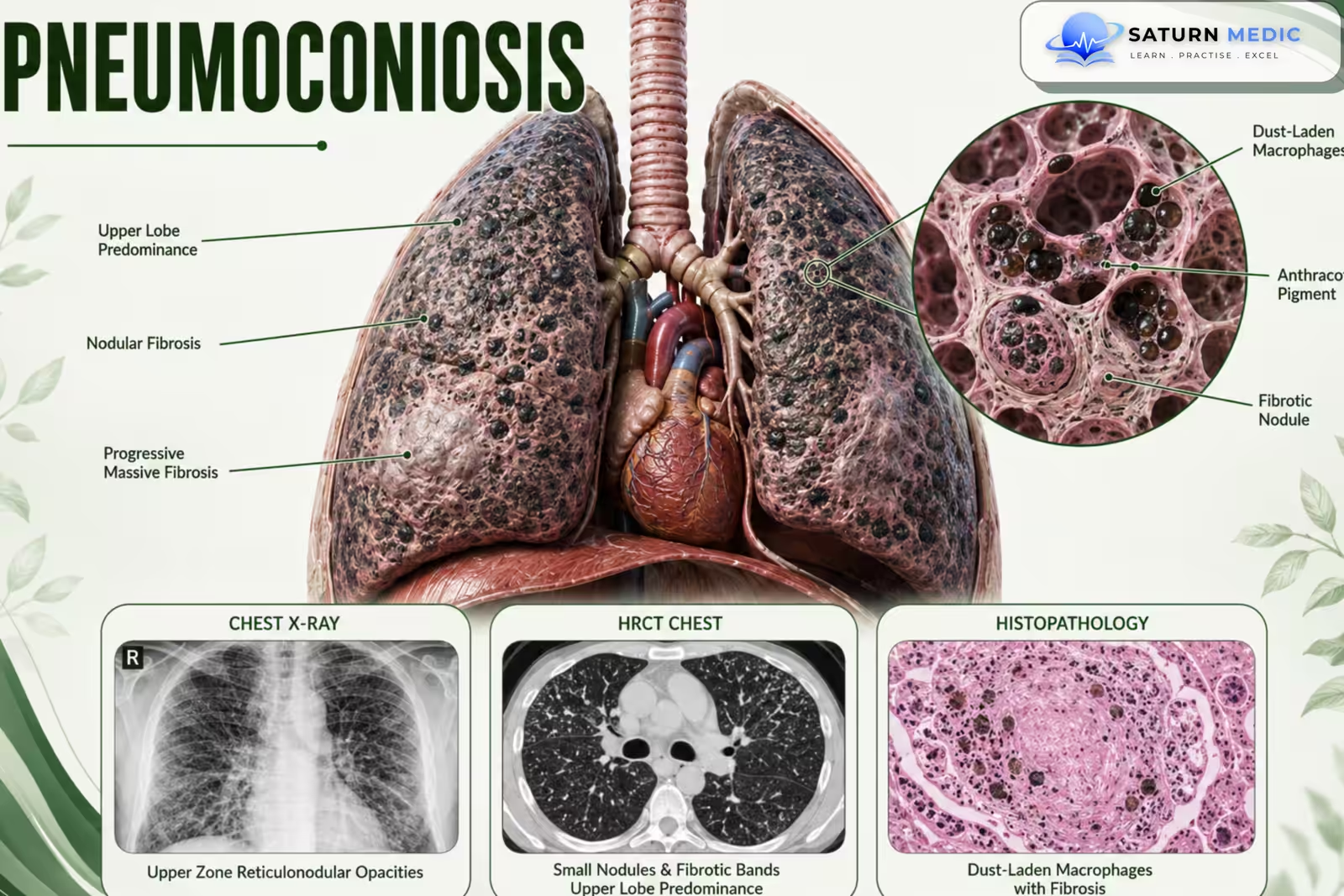
Pneumoconiosis is a group of occupational lung diseases caused by long-term inhalation and accumulation of inorganic dust particles in the lungs, leading to chronic inflammation and sometimes pulmonary fibrosis.
Also called
ICD-10
Specialty
Onset
Reviewed
-
OverviewOverview
-
Etiology & Risk FactorsEtiology & Risk Factors
-
PathophysiologyPathophysiology
-
Clinical PresentationClinical Presentation
-
History TakingHistory Taking
-
Physical ExaminationPhysical Examination
-
ClassificationClassification
-
InvestigationsInvestigations
-
DiagnosisDiagnosis
-
ManagementManagement
-
ComplicationsComplications
-
PrognosisPrognosis
-
Key Points / Clinical PearlsKey Points / Clinical Pearls
Overview
Pneumoconiosis develops after repeated exposure to harmful mineral dusts, usually in the workplace.
Major types include:
- Silicosis → Silica dust
- Coal workers’ pneumoconiosis (CWP) → Coal dust
- Asbestosis → Asbestos fibers
The disease may progress from dust deposition and inflammation to permanent pulmonary fibrosis.
Etiology & Risk Factors
-Main Causes
Silica Dust
Associated with:
Mining
Sandblasting
Stone cutting
Construction
Coal Dust
Associated with:
Coal mining
Asbestos Fibers
Associated with:
Construction
Shipbuilding
Insulation work
Demolition
Other Dusts
Beryllium
Talc
Iron
Other mineral dusts
Risk Factors
Long duration of exposure
High concentration of inhaled dust
Poor workplace ventilation
Lack of respiratory protection
Smoking
Certain high-risk occupations
Important Note
A detailed occupational history is essential in every patient with suspected pneumoconiosis.
Pathophysiology
Repeated inhalation of inorganic dust
⬇
Dust deposition in the lungs
⬇
Macrophage activation
⬇
Release of inflammatory mediators
⬇
Chronic lung inflammation
⬇
Fibroblast activation + Collagen deposition
⬇
Pulmonary fibrosis
⬇
Stiff lungs + Impaired gas exchange
⬇
Progressive dyspnea and respiratory impairment
Key Concept
The type of dust determines the pattern, severity, and complications of lung disease.
Clinical Presentation
-Symptoms
Progressive exertional dyspnea
Chronic cough
Sputum production in some patients
Chest tightness
Reduced exercise tolerance
Advanced Disease
Dyspnea at rest
Fatigue
Hypoxemia
Respiratory failure
-Signs
Tachypnea
Fine inspiratory crackles
Reduced oxygen saturation
Digital clubbing in some forms
Advanced Signs
Cyanosis
Signs of pulmonary hypertension
Signs of cor pulmonale
History Taking
Ask about:
- Shortness of breath?
- Chronic cough?
- When did symptoms start?
- Current occupation?
- Previous occupations?
- Mining work?
- Construction work?
- Stone cutting or sandblasting?
- Asbestos exposure?
- Duration of exposure?
Physical Examination
-General Examination
Look for:
- Tachypnea
- Cyanosis
- Reduced oxygen saturation
- Digital clubbing
-Chest Examination
May show:
- Fine inspiratory crackles
- Reduced breath sounds
- Signs of restrictive lung disease
-Advanced Disease
Look for:
- Raised JVP
- Peripheral edema
- Signs of pulmonary hypertension
- Signs of cor pulmonale
Important Note
Physical examination may be normal in early disease.
Classification
Occupational Lung Disease · Pneumoconiosis — All Types
Cause permanent fibrosis
Benign — no fibrosis
Most progress after cessation
Biopsy rarely needed
Exposure: Arc welding, iron foundries, iron ore mining, hematite mining
CXR: Dense nodular opacities (iron is radiopaque) — may appear dramatic but are benign
Key feature: No fibrosis, no functional impairment. Nodules may disappear after cessation. Welder’s lung — if coexisting silica or asbestos exposure, fibrosis can occur.
Benign — no fibrosis
Exposure: Tin mining, tin smelting, tin ore processing
CXR: Dense bilateral nodular opacities — very radiopaque (tin is dense). Striking appearance with normal or near-normal PFTs.
Key feature: No fibrosis, no symptoms, no functional impairment. Often discovered incidentally. One of the most radiologically dramatic but clinically benign pneumoconioses.
Benign — no fibrosis
Exposure: Barytes mining and milling, drilling mud manufacturing, rubber / paint industry
CXR: Very dense discrete nodules throughout both lung fields — extremely radiopaque. May be mistaken for metastases.
Key feature: No fibrosis, no symptoms, no functional impairment. PFTs normal. Benign incidental finding in exposed workers. Barium sulfate is inert.
Benign — no fibrosis
| Type | Dust | Occupation | CXR pattern | ILO opacity | PFT | Key complication | Fibrosis |
|---|---|---|---|---|---|---|---|
| Silicosis | Crystalline SiO₂ | Mining, sandblasting, stone cutting, engineered quartz | Rounded nodules, upper lobe; eggshell calcification; PMF | p/q/r (rounded) | Restrictive | TB (×4), lung cancer, PMF, scleroderma | Yes — severe |
| CWP | Coal dust | Underground coal mining | Rounded nodules, upper & mid zone; PMF; focal emphysema; no eggshell | p/q (rounded) | Restrictive / mixed | PMF, focal emphysema, Caplan syndrome | Yes |
| Asbestosis | Asbestos fibres | Shipbuilding, insulation, construction | Irregular opacities, lower lobe; pleural plaques; honeycombing | s/t (irregular) | Restrictive; ↓DLCO | Mesothelioma, lung cancer, pleural plaques, rounded atelectasis | Yes — lower lobe |
| Berylliosis (CBD) | Beryllium | Aerospace, nuclear, electronics | Hilar adenopathy; reticulonodular; identical to sarcoidosis | Variable | Restrictive; ↓DLCO | Granulomatous fibrosis; cor pulmonale | Yes — granulomatous |
| Byssinosis | Cotton / flax / hemp dust | Textile mills, cotton gins | Usually normal; hyperinflation late | N/A | Obstructive (unique) | Chronic airflow obstruction (COPD-like) | Minimal |
| Siderosis | Iron oxide | Welding, iron foundries | Dense nodules — radiopaque; dramatic but benign | p/q (rounded) | Normal | None (unless mixed with silica) | No |
| Stannosis | Tin oxide | Tin mining & smelting | Very dense radiopaque nodules; striking but benign | p/q (rounded) | Normal | None | No |
| Baritosis | Barium sulfate | Barytes mining, drilling | Very dense radiopaque nodules; may mimic metastases | p (rounded) | Normal | None | No |
Investigations
-Chest Imaging — Key Investigation
Chest X-ray
May show:
Small rounded opacities
Reticular changes
Pleural abnormalities
Large fibrotic masses in advanced disease
High-Resolution CT (HRCT)
Provides better assessment of:
Dust-related nodules
Fibrosis
Progressive massive fibrosis
Pleural disease
-Pulmonary Function Tests
May show:
Restrictive pattern
Obstructive pattern
Mixed pattern
Reduced DLCO
-Oxygen Assessment
Pulse oximetry
Exercise oxygen testing
Arterial blood gas in severe disease
-Additional Tests
Depending on the suspected type:
Tuberculosis testing
Bronchoscopy
Lung biopsy when the diagnosis remains uncertain
Diagnosis
Occupational Medicine · Pneumoconiosis — Diagnostic Criteria
Mining, sandblasting
Upper zones — nodular
Underground mining
Simple → PMF
Shipbuilding, insulation
Lower zones — fibrosis
Talcosis, Byssinosis
Dust-specific patterns
| Feature | Silicosis | CWP | Asbestosis | Berylliosis |
|---|---|---|---|---|
| Dust | Crystalline silica | Coal dust | Asbestos fibres | Beryllium |
| Latency | 10–30 yr | 10–20 yr | 20–40 yr | Months–decades |
| ILO opacity type | Rounded (p/q/r) | Rounded (p/q/r) | Irregular (s/t/u) | Nodular / granulomatous |
| CXR zone | Upper > lower | Upper > lower | Lower > upper | Upper > lower |
| Pathognomonic sign | Eggshell calcification | PMF masses | Asbestos bodies | BeLPT positive |
| Pleural disease | Rare | Rare | Plaques & thickening | Rare |
| PFT | Restrictive (PMF) | Restrictive ± obstructive | Restrictive, DLCO↓ | Restrictive, DLCO↓ |
| Malignancy | Lung Ca (IARC Gr1) | Minimal | Lung Ca, Mesothelioma | Lung Ca (possible) |
| TB risk | High (×3) | Moderate | Low | Low |
| Caplan syndrome | Yes | Yes (classic) | Yes | No |
Related Topics
Management
-Main Goals
Stop further exposure
Control symptoms
Preserve lung function
Prevent complications
-Exposure Control
Remove or reduce further dust exposure
Use appropriate respiratory protection
Improve workplace safety
-Supportive Management
Smoking cessation
Vaccination
Pulmonary rehabilitation
Oxygen therapy when indicated
Bronchodilators if airflow obstruction is present
-Treat Complications
Respiratory infections
Tuberculosis when present
Pulmonary hypertension
Respiratory failure
-Advanced Disease
Lung transplantation may be considered in selected patients
Important Note
Established pulmonary fibrosis is usually irreversible; prevention and early exposure control are essential.
Complications
- Progressive pulmonary fibrosis
- Progressive massive fibrosis
- Chronic hypoxemia
- Respiratory failure
- Pulmonary hypertension
- Cor pulmonale
- Tuberculosis
- Lung cancer in some dust exposures
- Mesothelioma with asbestos exposure
- Death
Prognosis
- Prognosis depends on:
- Type of inhaled dust
- Intensity of exposure
- Duration of exposure
- Extent of pulmonary fibrosis
- Presence of complications
- Mild disease may remain stable after exposure stops.
- Advanced fibrotic disease may continue to progress.
- Severe disease can lead to respiratory failure and pulmonary hypertension.
- Prevention of further exposure is essential for improving long-term outcomes.
Key Points / Clinical Pearls
- Pneumoconiosis is a group of occupational dust-related lung diseases.
- Silicosis, coal workers’ pneumoconiosis, and asbestosis are major types.
- A detailed occupational history is essential.
- Symptoms may appear years after exposure.
- Chest X-ray and HRCT are the main imaging tests.
- Pulmonary function may show restrictive, obstructive, or mixed abnormalities.
- Established fibrosis is usually irreversible.
- Stopping further exposure is the most important intervention.
- Silica exposure increases the risk of tuberculosis.
- Asbestos exposure increases the risk of lung cancer and mesothelioma.
- DeLight N, Sachs H. National Center for Biotechnology Information (NIH). Pneumoconiosis, StatPearls.
- Mlika M, Adigun R, Bhutta BS. National Center for Biotechnology Information (NIH). Silica-Induced Pneumoconiosis, StatPearls.
- Pathology and Mineralogy of the Pneumoconioses. PMC11617341.
- MedlinePlus, National Library of Medicine (NIH). Pneumoconiosis: Medical Encyclopedia.
- National Center for Biotechnology Information (NIH). Asbestosis, StatPearls.