

Clinical Subject Page
Tuberculosis (TB)
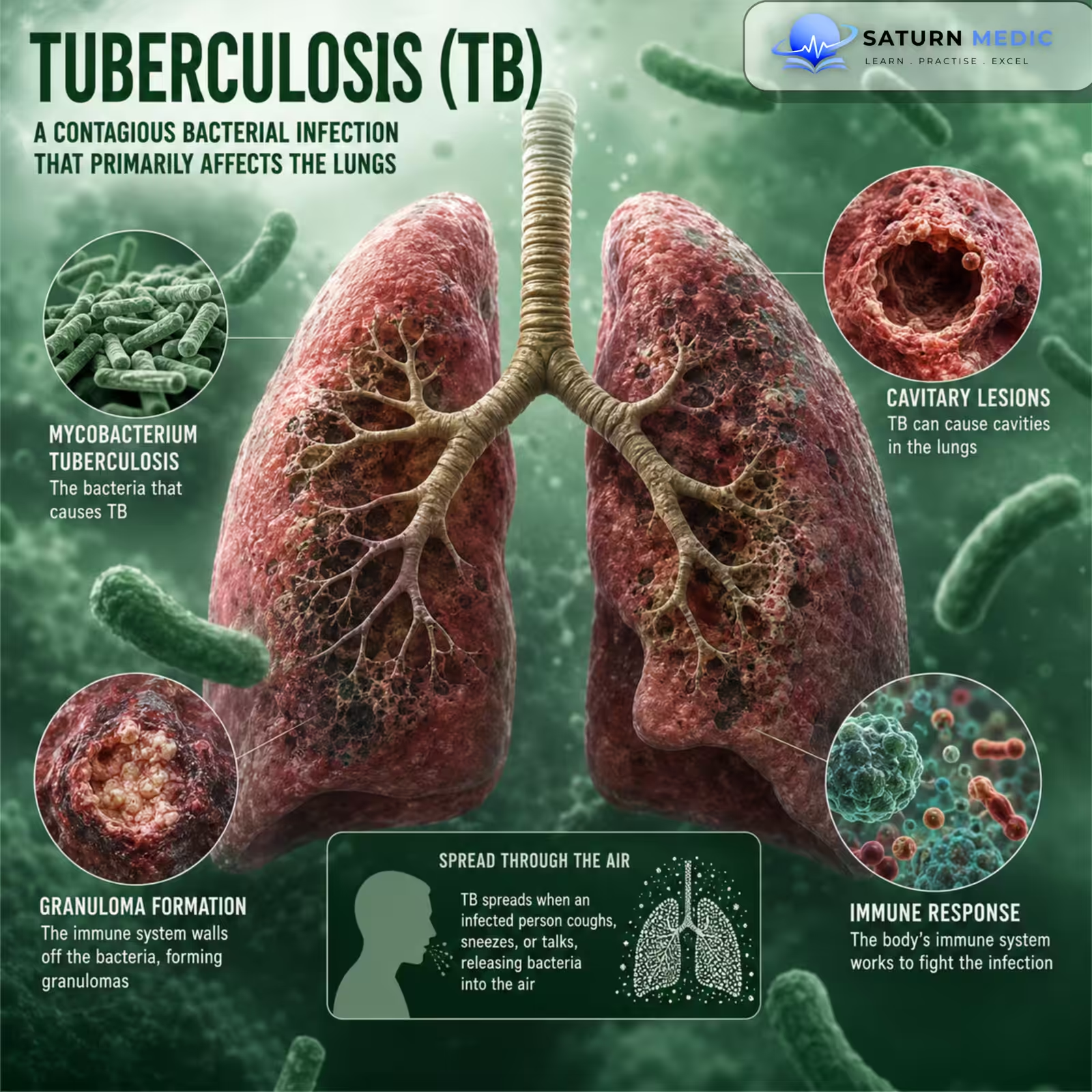
Tuberculosis (TB) is an infectious disease caused by Mycobacterium tuberculosis, most commonly affecting the lungs but capable of involving almost any organ.
Also called
ICD-10
Specialty
Onset
Reviewed
-
OverviewOverview
-
Etiology & Risk FactorsEtiology & Risk Factors
-
PathophysiologyPathophysiology
-
Clinical PresentationClinical Presentation
-
History TakingHistory Taking
-
Physical ExaminationPhysical Examination
-
InvestigationsInvestigations
-
DiagnosisDiagnosis
-
ManagementManagement
-
ComplicationsComplications
-
PrognosisPrognosis
-
Key Points / Clinical PearlsKey Points / Clinical Pearls
Overview
Tuberculosis spreads through the air when a person with infectious pulmonary or laryngeal Tuberculosis releases bacteria.
After infection, Tuberculosis may exist as:
- Latent Tuberculosis infection (LTBI) → Infection without active disease
- Active Tuberculosis disease → Bacterial multiplication causing symptoms and tissue damage
The lungs are the most commonly affected site.
Etiology & Risk Factors
-Cause
Tuberculosis is caused mainly by:
-
Mycobacterium tuberculosis
-Transmission
Spread occurs through airborne particles released by a person with infectious pulmonary or laryngeal Tuberculosis.
-Risk Factors for Exposure
-
Close contact with a person with active Tuberculosis
-
Living or working in crowded settings
-
Residence in areas with a high Tuberculosis burden
-
Healthcare exposure
-
Poor ventilation
Risk Factors for Active Disease
-
HIV infection
-
Immunosuppressive therapy
-
Malnutrition
-
Diabetes mellitus
-
Chronic kidney disease
-
Silicosis
-
Smoking
-
Young age
-
Older age
Important Note
Latent Tuberculosis infection is not contagious; active pulmonary or laryngeal Tuberculosis may be infectious.
Pathophysiology
Inhalation of Mycobacterium tuberculosis
⬇
Bacteria reach the alveoli
⬇
Macrophage uptake
⬇
Intracellular bacterial survival and multiplication
⬇
Cell-mediated immune response
⬇
Granuloma formation
⬇
Two Possible Outcomes
Immune control
→ Latent Tuberculosis infection
OR
Failure of immune control
→ Active Tuberculosis disease
→ Caseous necrosis + Tissue destruction
Key Concept
Tuberculosis may remain latent for years and later reactivate when immune control weakens.
Clinical Presentation
Pulmonary Symptoms
-
Persistent cough
-
Sputum production
-
Hemoptysis
-
Chest pain
-
Dyspnea in advanced disease
Constitutional Symptoms
-
Fever
-
Night sweats
-
Weight loss
-
Fatigue
-
Loss of appetite
Extrapulmonary Tuberculosis
May affect:
-
-
Lymph nodes
-
Pleura
-
Bones and joints
-
Central nervous system
-
Genitourinary system
-
Abdomen
-
Pericardium
-
History Taking
-Ask about:
-
- Persistent cough?
- How long has it lasted?
- Sputum production?
- Hemoptysis?
- Fever?
- Night sweats?
- Weight loss?
- Loss of appetite?
- Close contact with a person with Tuberculosis?
- Previous Tuberculosis infection or disease?
- Previous Tuberculosis treatment?
- Travel or residence in a high-burden area?
Physical Examination
-General Examination
Look for:
- Weight loss
- Fever
- Cachexia
- Pallor
- Lymphadenopathy
- Respiratory distress
-Chest Examination
May show:
- Crackles
- Bronchial breathing
- Reduced breath sounds
- Signs of pleural effusion
Important Note
Physical examination may be normal, especially in early pulmonary Tuberculosis.
-Extrapulmonary Examination
Look for:
- Enlarged lymph nodes
- Spinal tenderness or deformity
- Neurological abnormalities
- Abdominal swelling
- Signs of pericardial disease
Investigations of Tuberculosis
-Chest Imaging
Chest X-ray
May show:
-
Upper-lobe infiltrates
-
Cavitation
-
Nodules
-
Fibrosis
-
Hilar or mediastinal lymphadenopathy
-
Pleural effusion
CT Chest
Provides better assessment of:
-
Cavities
-
Nodules
-
Lymphadenopathy
-
Extent of disease
-Microbiological Testing — Essential for Active Tuberculosis
Sputum Tests
-
Nucleic acid amplification test (NAAT)
-
Acid-fast bacillus (AFB) smear
-
Mycobacterial culture
Key Point
-Mycobacterial culture confirms the organism and allows drug-susceptibility testing.
-Tests for Tuberculosis Infection
-
Tuberculin skin test (TST)
-
Interferon-gamma release assay (IGRA)
Important Note
-TST and IGRA detect Tuberculosis infection but cannot alone distinguish latent Tuberculosis from active Tuberculosis disease.
-Additional Tests
-
-
HIV testing
-
CBC
-
Liver function tests
-
Renal function tests
-
Testing of extrapulmonary specimens when indicated
-
Diagnosis
TB Diagnosis · Steps 1–2 — History & Examination
TB Diagnosis · Steps 3–4 — Imaging & Immunological Tests
| Test | Mechanism | Positive threshold | Advantages | Limitations |
|---|---|---|---|---|
| TST (Mantoux) Intradermal |
0.1 mL PPD injected. Read induration at 48–72h. | ≥5 mm HIV+, close contact ≥10 mm high-risk groups ≥15 mm low-risk |
Cheap, no lab. Useful in low-resource settings. | False positive with BCG. 2 visits. Anergy in immunosuppression. |
| IGRA QuantiFERON / T-SPOT Blood test |
Blood incubated with TB antigens (ESAT-6, CFP-10). Measures IFN-γ release. | Positive / Negative / Indeterminate | Not affected by BCG. Single visit. Preferred in BCG-vaccinated. | Expensive. Indeterminate in severe immunosuppression. |
TB Diagnosis · Steps 5–6 — Microbiology & Extra-pulmonary Workup
CSF Xpert — sensitivity ~60%; test of choice
Brain MRI — basilar enhancement, tuberculomas, hydrocephalus
Pleural biopsy — highest yield (~80%)
Fluid: exudate, lymphocyte-predominant, low glucose
Bone marrow biopsy — miliary TB
Urine AFB × 3 — renal TB (early morning urine)
Related Topics
Management of Tuberculosis
TB Treatment · Standard First-line Regimen (DS-TB) — WHO 2022
| Drug (abbreviation) | Daily dose (adult) | Phase | Duration | Principal side effects | Monitoring |
|---|---|---|---|---|---|
|
Isoniazid
H
5 mg/kg/day
(max 300 mg) |
300 mg OD Fasting or with food |
Intensive Continuation |
6 months |
Peripheral neuropathy
Hepatotoxicity
Drug-induced lupus
Mood changes
Rash
Neuropathy prevented by pyridoxine (B6) 10–25 mg/day — give routinely to HIV+, malnourished, diabetics, alcohol users, pregnancy
|
LFTs at baseline & if symptomatic
Symptoms of neuropathy
|
|
Rifampicin
R
10 mg/kg/day
(max 600 mg) |
600 mg OD 30 min before food |
Intensive Continuation |
6 months |
Hepatotoxicity
Orange discolouration
Flu-like syndrome
Drug interactions
Thrombocytopenia
Potent CYP450 inducer — reduces efficacy of OCP, warfarin, antiretrovirals, phenytoin, methadone. Warn patients: urine/sweat/tears turn orange — stains contact lenses permanently.
|
LFTs at baseline
Drug interaction review
FBC if prolonged
|
|
Pyrazinamide
Z
25 mg/kg/day
(max 2g) |
1.5–2 g OD With food |
Intensive only | 2 months |
Hepatotoxicity
Hyperuricaemia / gout
Arthralgia
Rash, nausea
Photosensitivity
Asymptomatic hyperuricaemia common — treat only if symptomatic gout. Avoid in severe hepatic disease and gout.
|
LFTs at baseline & monthly
Uric acid if symptomatic
Joint symptoms
|
|
Ethambutol
E
15–20 mg/kg/day
(max 1.6 g) |
800–1200 mg OD With or without food |
Intensive only | 2 months |
Optic neuritis
Colour vision loss (red-green)
Peripheral neuropathy
Rash
Optic neuritis is dose-dependent and usually reversible if caught early — warn patients to report blurred vision or colour change immediately. Avoid in pre-existing optic nerve disease. Dose-reduce in renal impairment.
|
Visual acuity at baseline
Red-green colour test
Monthly symptoms check
eGFR (dose-adjust if low)
|
Extended to 9 months (2HRZE / 7HR) for: CNS TB (meningitis), bone/joint TB, extensive pulmonary disease, HIV-positive patients with CD4 <100.
All four drugs are given together in fixed-dose combination (FDC) tablets where available — improves adherence and reduces risk of inadvertent monotherapy.
TB Treatment · MDR-TB Regimen BPaLM / BPaL — WHO 2022
| Drug (abbreviation) | Daily dose (adult) | Phase | Duration | Principal side effects | Monitoring |
|---|---|---|---|---|---|
|
Bedaquiline
B
400 mg OD × 2 weeks
then 200 mg 3×/week |
With food (increases absorption) |
All phases | 6 months |
QTc prolongation
Hepatotoxicity
Nausea, arthralgia
Headache
Avoid with other QT-prolonging drugs. Do NOT combine with fluoroquinolones without ECG monitoring. Diarylquinoline class — novel mechanism (inhibits ATP synthase).
|
ECG at baseline, 2, 12, 24 weeks
LFTs monthly
Serum K, Mg, Ca
|
|
Pretomanid
Pa
200 mg OD
|
With food | All phases | 6 months |
Peripheral neuropathy
Hepatotoxicity
QTc prolongation
Anaemia, rash
Nitroimidazooxazine class — requires reductive activation by bacillary F420 enzyme. Only used within BPaL/BPaLM regimen.
|
LFTs monthly
Neuropathy symptoms
ECG monitoring
FBC
|
|
Linezolid
L
600 mg OD
(reduce to 300 mg if toxicity) |
OD — may reduce dose for tolerability | All phases | 6 months |
Myelosuppression
Peripheral neuropathy
Optic neuropathy
Serotonin syndrome
Nausea, diarrhoea
Most toxic drug in regimen. Dose reduction to 300 mg OD often required. Avoid SSRIs, MAOIs (serotonin syndrome). Monitor FBC weekly initially.
|
FBC weekly × 4, then monthly
Visual acuity monthly
Neuropathy assessment
Drug interactions review
|
|
Moxifloxacin
M
400 mg OD
|
OD — added in BPaLM if susceptible | BPaLM only | 6 months |
QTc prolongation
Tendinopathy / tendon rupture
Arthralgia, myalgia
GI upset, dizziness
Photosensitivity
Added only if moxifloxacin-susceptible strain confirmed. Additive QTc risk with bedaquiline — mandatory ECG monitoring. Avoid in fluoroquinolone-resistant MDR-TB.
|
ECG at baseline & monthly
Tendon pain symptoms
Serum electrolytes
|
BPaLM regimen (preferred, 2022): Bedaquiline + Pretomanid + Linezolid + Moxifloxacin × 6 months — if moxifloxacin-susceptible.
BPaL regimen: Bedaquiline + Pretomanid + Linezolid × 6–9 months — if fluoroquinolone-resistant (XDR-TB).
Confirm DST before starting. Treat under DOTS (directly observed therapy) supervision. Notify TB authorities. All MDR-TB managed by specialist TB unit.
TB Treatment · Latent TB Infection (LTBI) Preventive Therapy — WHO 2022
| Regimen | Dosing | Schedule | Duration | Principal side effects | Monitoring |
|---|---|---|---|---|---|
|
Isoniazid alone
6H
5 mg/kg/day (max 300 mg)
|
300 mg OD | Daily | 6 months |
Hepatotoxicity
Peripheral neuropathy
Rash, GI upset
Give pyridoxine 10–25 mg/day with all INH-containing LTBI regimens. Hepatotoxicity risk increases with age and alcohol use.
|
LFTs at baseline
Monthly symptom review
Adherence assessment
|
|
Isoniazid alone
9H
5 mg/kg/day (max 300 mg)
|
300 mg OD | Daily | 9 months |
Hepatotoxicity
Peripheral neuropathy
Rash, GI upset
Preferred by CDC guidelines. More protective than 6H (~90% efficacy). Higher hepatotoxicity risk with longer duration. Preferred for HIV-positive patients when rifampicin interactions are a concern.
|
LFTs at baseline & if symptomatic
Monthly review
|
|
Rifampicin alone
4R
10 mg/kg/day (max 600 mg)
|
600 mg OD | Daily | 4 months |
Hepatotoxicity
Drug interactions (CYP inducer)
Orange discolouration
Flu-like reactions
BTS-preferred regimen for LTBI. Fewer side effects and better completion rates than 6H. Lower hepatotoxicity. Check drug interactions (OCP, antiretrovirals, warfarin).
|
LFTs at baseline
Drug interaction review
Monthly adherence review
|
|
Rifampicin + Isoniazid
3HR
Rifampicin 600 mg + Isoniazid 300 mg OD
|
Both OD together 30 min before food |
Daily | 3 months |
Hepatotoxicity (additive)
Peripheral neuropathy
Drug interactions
GI upset, rash
Widely used WHO-endorsed short regimen. Good adherence due to shorter duration. Give pyridoxine. Monitor LFTs baseline + monthly.
|
LFTs baseline & monthly
Drug interactions
Pyridoxine prescribed
|
|
Rifapentine + Isoniazid
3HP
Rifapentine weight-based + INH 900 mg
|
Once weekly — DOT or self-administered | Weekly | 3 months (12 doses) |
Hepatotoxicity
Flu-like reactions (systemic)
Drug interactions
Rash, hypersensitivity
Highest completion rates of all LTBI regimens. Preferred by CDC 2020. Once-weekly dosing ideal for DOT. Not recommended in HIV+ on antiretrovirals (major drug interactions). Avoid in pregnancy and age <2 years.
|
LFTs at baseline
Systemic reaction symptoms
Drug interactions (HIV ARVs)
|
Priority groups for LTBI treatment: HIV-positive, close contacts of smear-positive TB, immunosuppressed (anti-TNF, transplant, steroids), new migrants from high-burden countries, those with fibrotic CXR changes (old healed TB).
Pyridoxine 10–25 mg/day with all isoniazid-containing regimens to prevent peripheral neuropathy.
Complications of Tuberculosis
- Massive hemoptysis
- Permanent lung damage
- Bronchiectasis
- Respiratory failure
- Pleural disease
- Disseminated or miliary Tuberculosis
- Tuberculosis meningitis
- Spinal Tuberculosis
- Pericardial Tuberculosis
- Drug-resistant Tuberculosis
- Death
Prognosis of Tuberculosis
- Drug-susceptible Tuberculosis is usually curable with appropriate treatment.
- Prognosis depends on:
- Early diagnosis
- Drug susceptibility
- Treatment adherence
- Disease extent
- Immune status
- Presence of complications
- Delayed diagnosis increases transmission and complications.
- Drug-resistant Tuberculosis is more difficult to treat and generally requires longer, more complex therapy.
- Early diagnosis and completion of treatment greatly improve outcomes.
Key Points / Clinical Pearls of Tuberculosis
- Tuberculosis is caused mainly by Mycobacterium tuberculosis.
- Pulmonary Tuberculosis is the most common form.
- Latent Tuberculosis infection is not contagious.
- Active pulmonary or laryngeal Tuberculosis may be infectious.
- Persistent cough, weight loss, fever, and night sweats are classic features.
- NAAT, AFB smear, and culture are important tests for active pulmonary Tuberculosis.
- TST and IGRA cannot alone distinguish latent from active Tuberculosis.
- Always exclude active disease before treating latent Tuberculosis infection.
- Tobin EH, Tristram D. National Center for Biotechnology Information (NIH). Tuberculosis Overview, StatPearls.
- World Health Organization. Tuberculosis: Fact Sheet.
- Price C, Nguyen AD. National Center for Biotechnology Information (NIH). Latent Tuberculosis, StatPearls.
- Swinkels HM, Jilani TN, Tobin EH. National Center for Biotechnology Information (NIH). Tuberculosis Prevention, Control, and Elimination, StatPearls.
- World Health Organization. Global Tuberculosis Report 2025.