

Clinical Subject Page
Posterior Urethral Valves (PUV)
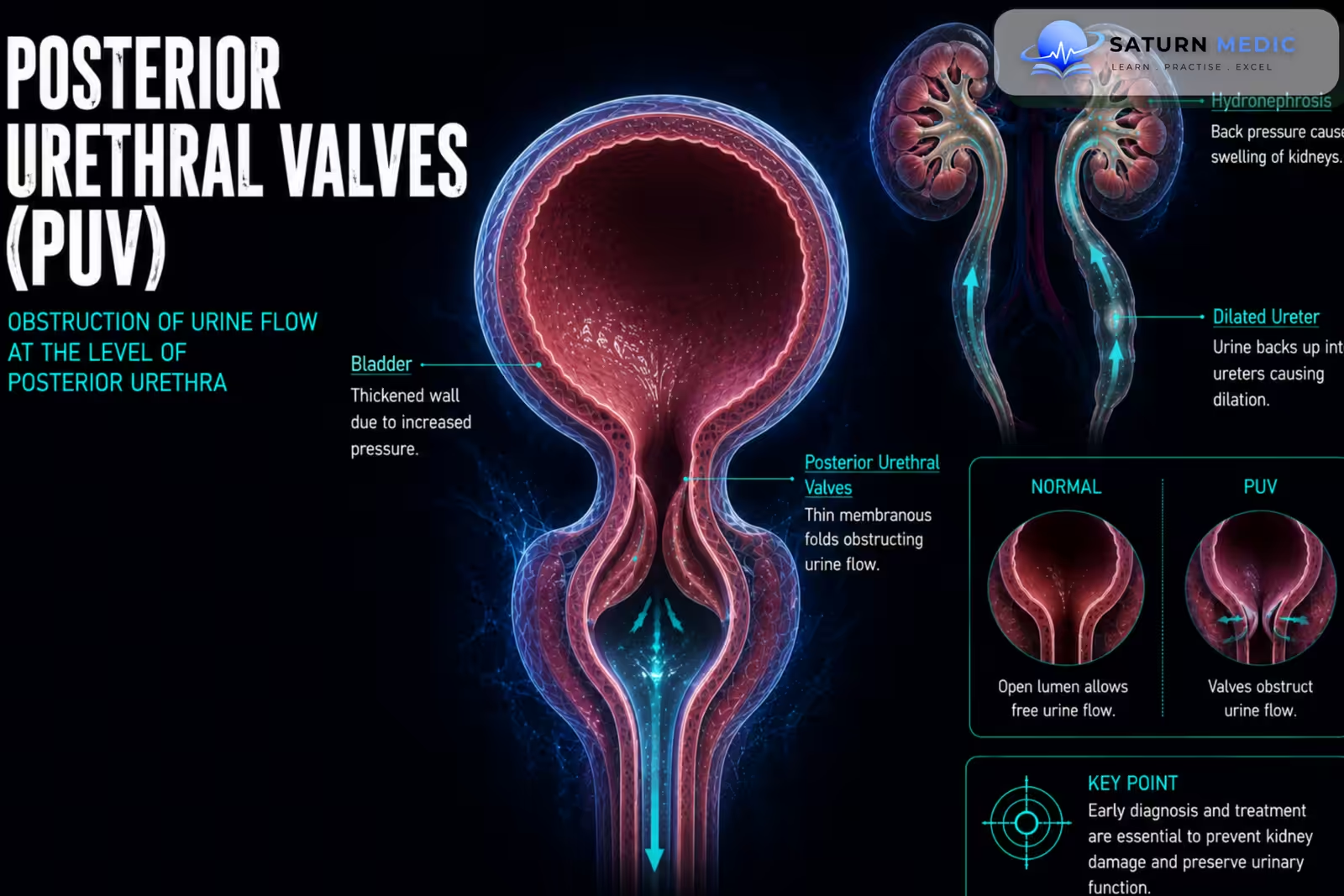
Posterior Urethral Valves (PUV) are congenital obstructing membranous folds within the posterior urethra that occur only in males. They cause bladder outlet obstruction and can lead to progressive damage to the bladder, ureters, and kidneys
Also called
ICD-10
Specialty
Onset
Reviewed
-
OverviewOverview
-
Etiology & Risk FactorsEtiology & Risk Factors
-
PathophysiologyPathophysiology
-
Clinical PresentationClinical Presentation
-
History TakingHistory Taking
-
Physical ExaminationPhysical Examination
-
InvestigationsInvestigations
-
DiagnosisDiagnosis
-
ManagementManagement
-
ComplicationsComplications
-
PrognosisPrognosis
-
Key Points / Clinical PearlsKey Points / Clinical Pearls
Overview
-Posterior Urethral Valves (PUV) are an important cause of lower urinary tract obstruction in male infants and children.
-Obstruction causes increased pressure within the urinary tract, leading to:
- Bladder wall thickening and dysfunction
- Hydroureter
- Hydronephrosis
- Vesicoureteral Reflux (VUR)
- Renal dysplasia
- Progressive Chronic Kidney Disease CKD
-The severity ranges from mild obstruction diagnosed later in childhood to severe bilateral urinary tract damage detected before birth
Etiology & Risk Factors
-Etiology
–Posterior Urethral Valves (PUV) result from abnormal embryological development of the posterior urethra, producing obstructing membranous folds that impair urinary outflow.
The condition is congenital and occurs exclusively in males.
-Risk Factors
- Male sex
- Congenital urinary tract abnormalities
- Antenatal ultrasound showing urinary tract obstruction
Pathophysiology
Posterior urethral obstruction → increased bladder outlet pressure → bladder distension and hypertrophy → increased pressure transmitted to ureters and kidneys → hydroureteronephrosis ± Vesicoureteral Reflux (VUR) → renal dysplasia and nephron damage → reduced kidney function → Chronic Kidney Disease CKD
Clinical Presentation
–Symptoms
-
Presentation depends on severity and age.
Possible symptoms include:
- Poor urinary stream
- Straining to void
- Dribbling
- Reduced urine output
- Recurrent urinary tract infections
- Urinary retention
- Failure to thrive
-Signs:
- Palpable distended bladder
- Poor urinary stream
- Enlarged kidneys in severe cases
- Failure to thrive
- Hypertension
History Taking
-Ask about:
- Antenatal ultrasound findings
- Oligohydramnios
- Poor urinary stream
- Straining or dribbling during urination
- Reduced urine output
- Urinary retention
- Recurrent urinary tract infections
- Fever
- Poor feeding
- Poor weight gain
Physical Examination
-General Examination
- Hydration status
- Weight and growth
- Blood pressure
- Signs of Chronic Kidney Disease CKD
-System-Specific Examination:
- Palpate for a distended bladder
- Assess abdominal masses from hydronephrotic kidneys
- Observe the urinary stream when possible
- Assess for signs of urinary tract infection
Investigations
-Biochemistry / Specific Tests
-
Serum creatinine to assess kidney function
-
Electrolytes, especially potassium and bicarbonate
-
Urinalysis
-
Urine culture when infection is suspected
-Imaging
Renal and Bladder Ultrasound
Used to assess:
-
Hydronephrosis
-
Hydroureter
-
Bladder distension
-
Bladder wall thickening
-
Renal size and appearance
-Voiding Cystourethrogram (VCUG)
The key diagnostic imaging investigation.
It may demonstrate:
-
Dilated posterior urethra
-
Abrupt narrowing distal to the obstruction
-
Thick-walled bladder
-
Vesicoureteral Reflux (VUR)
-The characteristic appearance may resemble a “keyhole sign”.
-Special / Confirmatory Tests
Cystoscopy directly confirms Posterior Urethral Valves (PUV) and allows simultaneous treatment by valve ablation.
Diagnosis
–Diagnosis of Posterior Urethral Valves (PUV) is based on:
Clinical suspicion in a male child + evidence of bladder outlet obstruction on ultrasound and VCUG + cystoscopic confirmation.
-Important diagnostic findings include:
- Dilated posterior urethra
- Thick-walled bladder
- Bilateral hydroureteronephrosis
- Vesicoureteral Reflux (VUR)
Management
1. First-Line / Emergency Management
Immediate priorities are to relieve urinary obstruction and stabilize the child.
-
Bladder drainage with an appropriately placed urinary catheter
-
Correct dehydration and electrolyte abnormalities
-
Treat urinary tract infection
-
Urgent pediatric urology involvement
2. Definitive Treatment
Endoscopic valve ablation is the definitive treatment in most patients.
-The obstructing valves are destroyed using
3. Medical Treatment
Medical management may include:
-
Antibiotics for urinary tract infection
-
Antibiotic prophylaxis in selected patients
-
Treatment of electrolyte abnormalities
-
Management of hypertension
-
Treatment of complications of Chronic Kidney Disease CKD
4. Surgical / Procedural Treatment
-
Urinary catheter drainage
-
Endoscopic valve ablation
-
Vesicostomy in selected infants when immediate ablation is not feasible
-
Upper urinary tract diversion in rare selected cases
-
Renal replacement therapy for severe End Stage Renal Disease ESRD
5. Supportive Management
-
Regular monitoring of kidney function
-
Blood pressure monitoring
-
Monitoring growth and nutrition
-
Assessment of bladder function
Complications
- Bladder dysfunction
- Vesicoureteral Reflux (VUR)
- Recurrent urinary tract infections
- Hydroureteronephrosis
- Renal dysplasia
- Hypertension
- Electrolyte abnormalities
- Chronic Kidney Disease CKD
- End Stage Renal Disease ESRD
Prognosis
The prognosis of Posterior Urethral Valves (PUV) depends mainly on the severity of obstruction, degree of renal dysplasia, kidney function at presentation, and long-term bladder function. Early diagnosis and relief of obstruction improve outcomes, but some children develop progressive Chronic Kidney Disease CKD despite successful valve ablation because kidney damage may have occurred before treatment
Key Points / Clinical Pearls
- Posterior Urethral Valves (PUV) are a congenital cause of bladder outlet obstruction in males.
- They can be detected before birth or present during infancy and childhood.
- Poor urinary stream is an important clinical clue.
- Severe obstruction can cause bilateral hydroureteronephrosis.
- The bladder may become thick-walled and dysfunctional.
- Renal and bladder ultrasound is an important initial investigation.
- Voiding cystourethrogram (VCUG) demonstrates posterior urethral obstruction and associated Vesicoureteral Reflux (VUR).
- Cystoscopy confirms the diagnosis and allows treatment.
- Initial management includes urgent bladder drainage and stabilization.
- Endoscopic valve ablation is the definitive treatment in most patients.
- European Association of Urology (EAU). EAU Guidelines on Paediatric Urology .
- National Institute of Diabetes and Digestive and Kidney Diseases (NIDDK). Information on Pediatric Urinary Tract and Urologic Disorders .
- Parkhouse HF, Woodhouse CRJ. Long-Term Status of Patients With Posterior Urethral Valves. Urol Clin North Am. 1990;17(2):373-378.
- Ansari MS, Gulia A, Srivastava A, Kapoor R. Risk Factors for Progression to End-Stage Renal Disease in Children With Posterior Urethral Valves. J Pediatr Urol. 2010;6(6):609-613. Journal of Pediatric Urology .
- Lopez Pereira P, Martinez Urrutia MJ, Espinosa L, et al. Bladder Dysfunction as a Prognostic Factor in Patients With Posterior Urethral Valves. BJU Int. 2002;90(3):308-311.
- National Library of Medicine (NIH). Posterior Urethral Valves . StatPearls.