

Clinical Subject Page
Nephrolithiasis
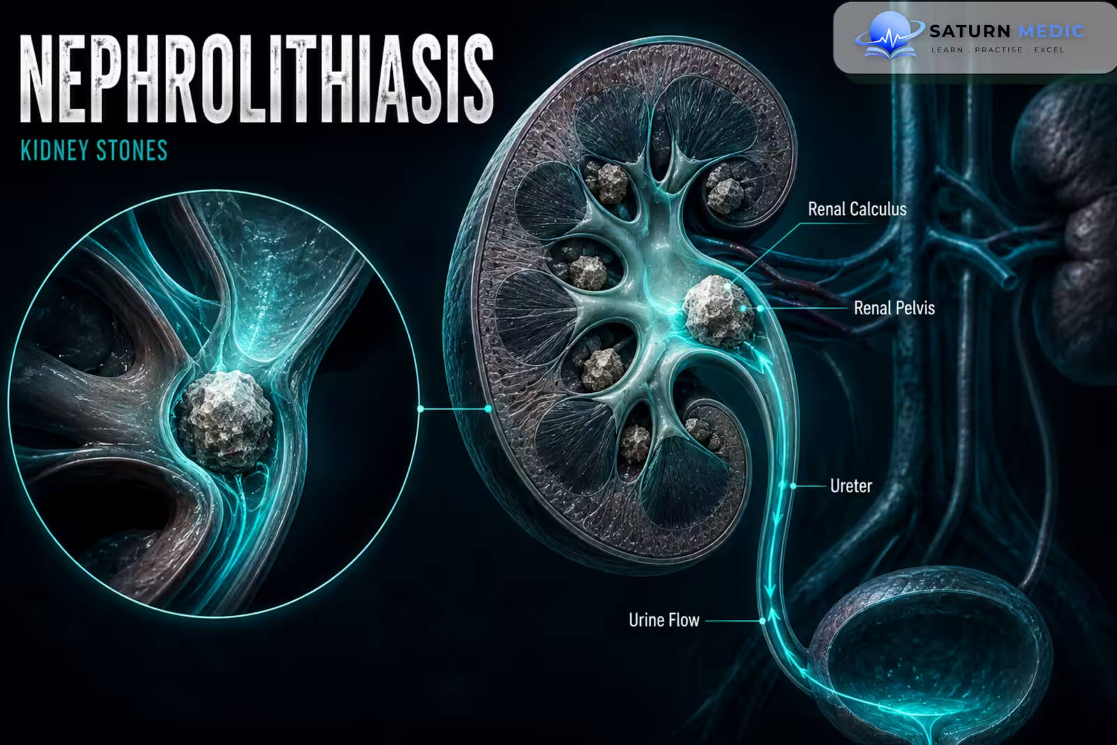
Nephrolithiasis is the formation of stones (calculi) within the kidneys or urinary tract. Stones may cause severe flank pain, hematuria, urinary obstruction, infection, and, in severe cases, Acute Kidney Injury (AKI)
Also called
ICD-10
Specialty
Onset
Reviewed
-
OverviewOverview
-
Etiology & Risk FactorsEtiology & Risk Factors
-
PathophysiologyPathophysiology
-
Clinical PresentationClinical Presentation
-
History TakingHistory Taking
-
Physical ExaminationPhysical Examination
-
InvestigationsInvestigations
-
DiagnosisDiagnosis
-
ManagementManagement
-
ComplicationsComplications
-
PrognosisPrognosis
-
Key Points / Clinical PearlsKey Points / Clinical Pearls
Overview
-Nephrolithiasis occurs when substances in urine become supersaturated and crystallize.
-The major stone types are:
- Calcium oxalate — most common
- Calcium phosphate
- Uric acid
- Struvite
- Cystine
-Clinical severity depends mainly on the stone’s size, location, degree of obstruction, and presence of infection.
Etiology & Risk Factors
-Etiology
–Stone formation results from urinary supersaturation, crystal formation, and inadequate inhibition of crystallization.
-Important causes include:
- Low urine volume
- Hypercalciuria
- Hyperoxaluria
- Hyperuricosuria
- Hypocitraturia
- Urinary Tract Infection with urease-producing organisms
- Genetic disorders such as cystinuria
-Risk Factors for Nephrolithiasis
- Low fluid intake
- Previous kidney stones
- High dietary sodium intake
- Obesity
- Gout or hyperuricemia
- Recurrent urinary tract infection
- Family history of stone disease
Pathophysiology
Urinary supersaturation → crystal nucleation → crystal growth and aggregation → stone formation → urinary tract obstruction or mucosal irritation → renal colic + hematuria ± infection
Clinical Presentation
-Symptoms:
- Sudden severe colicky flank pain
- Pain radiating to the groin or lower abdomen
- Hematuria
- Nausea and vomiting
- Dysuria
- Urinary frequency or urgency, particularly with distal ureteric stones
-Signs:
- Restlessness due to severe pain
- Costovertebral angle tenderness
- Flank tenderness
- Hematuria
History Taking
-Ask about:
- Character and location of pain
- Radiation of pain
- Previous kidney stones
- Hematuria
- Dysuria, frequency, or urgency
- Fever or chills
- Nausea and vomiting
- Fluid intake
- Dietary habits
- Family history of kidney stones
- History of gout
- Recurrent urinary tract infections
Physical Examination
-General Examination
- Temperature
- Blood pressure
- Hydration status
- Degree of pain or distress
-System-Specific Examination:
- Costovertebral angle tenderness
- Flank and abdominal tenderness
- Assessment for signs of urinary obstruction or sepsis
Investigations
-Biochemistry / Specific Tests
-
Urinalysis
-
Urine microscopy
-
Urine culture when infection is suspected
-
Serum creatinine and eGFR
-
Serum electrolytes
-
Serum calcium when evaluating recurrent stones
-
Serum uric acid when clinically indicated
-For recurrent or high-risk stone formers:
-
24-hour urine volume
-
Urinary calcium
-
Urinary oxalate
-
Urinary citrate
-
Urinary uric acid
-
Urinary sodium
-
Urine pH
-Imaging
Non-contrast CT of the kidneys, ureters, and bladder (CT KUB) is the most accurate imaging test for most adults with suspected acute stones.
Ultrasound is useful when radiation should be avoided, particularly in pregnancy, and can detect hydronephrosis.
Plain abdominal radiography may be useful for follow-up of selected radiopaque stones.
-Special / Confirmatory Tests
Stone analysis should be performed when a stone is retrieved, particularly in recurrent stone disease.
Metabolic evaluation is important in recurrent or high-risk patients.
Diagnosis
-Diagnosis Of Nephrolithiasis is based on:
- Typical renal colic
- Hematuria or supportive urinary findings
- Imaging confirmation of a urinary tract stone
Non-contrast CT KUB is the most accurate diagnostic test for most adults.
Urine culture is important when infection is suspected
Management
1. First-Line / Emergency Management
For acute renal colic:
-
NSAIDs are usually first-line analgesia when not contraindicated
-
Antiemetics for nausea and vomiting
-
Appropriate hydration according to clinical status
–Urgent intervention is required for:
-
Obstructed infected kidney
-
Sepsis
-
Anuria
-
Significant Acute Kidney Injury (AKI)
-
Solitary kidney with obstruction
-
Uncontrolled pain or persistent vomiting
2. Definitive Treatment
Management depends on stone size, location, composition, and likelihood of spontaneous passage.
-
Small ureteric stones may pass spontaneously
-
Larger or obstructing stones may require intervention
-
Stone prevention requires treatment of the underlying metabolic cause when identified
3. Medical Treatment
-
NSAIDs for renal colic when appropriate
-
Antiemetics
-
Alpha-blockers may be considered for selected ureteric stones to facilitate passage
-
Antibiotics when urinary infection is present
-
Potassium citrate for hypocitraturia and selected uric acid or cystine stones
-
Allopurinol for selected patients with recurrent hyperuricosuric calcium stones or uric acid stone disease
4. Surgical / Procedural Treatment
Depending on the stone:
-
Extracorporeal shock wave lithotripsy (ESWL)
-
Ureteroscopy with stone fragmentation or extraction
-
Percutaneous nephrolithotomy (PCNL)
-
Ureteric stent
-
Percutaneous nephrostomy
Infected obstruction requires urgent drainage with a ureteric stent or percutaneous nephrostomy.
5. Supportive Management
-
Increase fluid intake to maintain adequate urine volume
-
Reduce excessive dietary sodium
-
Maintain normal dietary calcium intake
-
Modify diet according to stone type
-
Maintain healthy body weight
-
Analyze retrieved stones
Complications
- Urinary tract obstruction
- Hydronephrosis
- Urinary tract infection
- Pyelonephritis
- Urosepsis
- Acute Kidney Injury (AKI)
- Recurrent stone formation
- Chronic Kidney Disease (CKD)
Prognosis
-The prognosis of Nephrolithiasis is generally good with appropriate treatment, and many small stones pass spontaneously.
-However, recurrence is common without preventive measures. Recurrent stones, persistent obstruction, or repeated infection can cause progressive renal damage and contribute to Chronic Kidney Disease (CKD).
Key Points / Clinical Pearls
- Nephrolithiasis means stone formation within the kidneys or urinary tract.
- Calcium oxalate is the most common type of urinary stone.
- Low urine volume is a major modifiable risk factor.
- Severe colicky flank pain radiating to the groin is a classic presentation.
- Hematuria commonly occurs.
- Fever with an obstructing stone suggests a potentially life-threatening infection.
- Urinalysis is an important initial investigation.
- Non-contrast CT KUB is the most accurate imaging test for most adults.
- Ultrasound is useful when radiation should be avoided.
- NSAIDs are usually first-line treatment for acute renal colic when appropriate.
- Small stones may pass spontaneously.
- Larger or obstructing stones may require ureteroscopy, ESWL, or PCNL.
- European Association of Urology (EAU). EAU Guidelines on Urolithiasis .
- Assimos D, Krambeck A, Miller NL, et al. Surgical Management of Stones: American Urological Association/Endourological Society Guideline. J Urol. 2016;196(4):1153-1160. Journal of Urology .
- Pearle MS, Goldfarb DS, Assimos DG, et al. Medical Management of Kidney Stones: AUA Guideline. J Urol. 2014;192(2):316-324. Journal of Urology .
- Khan SR, Pearle MS, Robertson WG, et al. Kidney Stones. Nat Rev Dis Primers. 2016;2:16008. Nature Reviews Disease Primers .
- National Institute of Diabetes and Digestive and Kidney Diseases (NIDDK). Kidney Stones . National Institutes of Health.
- National Library of Medicine (NIH). Urolithiasis . StatPearls.