
Clinical Subject Page

Peyronie's Disease
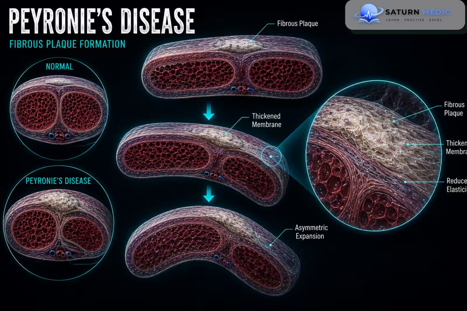
Peyronie’s Disease is an acquired condition in which fibrous scar tissue, called a plaque, develops in the tunica albuginea of the penis. This can cause penile curvature, shortening, deformity, pain during erection, and Erectile Dysfunction (ED)
Also called
ICD-10
Specialty
Onset
Reviewed
-
OverviewOverview
-
Etiology & Risk FactorsEtiology & Risk Factors
-
PathophysiologyPathophysiology
-
Clinical PresentationClinical Presentation
-
History TakingHistory Taking
-
Physical ExaminationPhysical Examination
-
InvestigationsInvestigations
-
DiagnosisDiagnosis
-
ManagementManagement
-
ComplicationsComplications
-
PrognosisPrognosis
-
Key Points / Clinical PearlsKey Points / Clinical Pearls
Overview
–Peyronie’s Disease results from localized fibrosis of the tunica albuginea. During erection, the affected area cannot expand normally, causing the penis to bend toward the side of the plaque.
The disease has two main phases:
- Active phase: penile pain and progressive changes in curvature or deformity.
- Stable phase: pain usually improves, and the deformity becomes stable.
Etiology & Risk Factors
-Etiology
–The exact cause is not fully understood. Repeated minor trauma to the erect penis is thought to cause abnormal wound healing, inflammation, and excessive collagen deposition, resulting in fibrous plaque formation.
-Risk Factors
- Penile trauma or repeated microtrauma
- Increasing age
- Erectile Dysfunction (ED)
- Diabetes Mellitus
- Dupuytren’s contracture
Pathophysiology
Penile microtrauma → inflammation of the tunica albuginea → abnormal wound healing → excessive collagen deposition → fibrous plaque formation → reduced tunical elasticity → asymmetric expansion during erection → penile curvature and deformity
Clinical Presentation
–Symptoms
- New penile curvature during erection
- Penile shortening
- Penile narrowing or indentation
- Hourglass deformity
- Painful erections, especially during the active phase
- Difficulty with sexual intercourse
- Erectile Dysfunction (ED)
-Signs:
- Palpable firm plaque within the penile shaft
- Curvature during erection
- Penile indentation or narrowing
- Reduced penile length
History Taking
-Ask about:
- When the curvature first appeared
- Whether the curvature is progressing
- Penile pain
- Presence of a palpable lump or plaque
- Degree and direction of curvature
- Penile shortening
- Difficulty with intercourse
- Erectile function
- Previous penile trauma
Physical Examination
-General Examination
- Assess for associated fibrotic disorders when clinically relevant
- Assess the psychological impact of the condition
-System-Specific Examination:
- Palpate the penis for fibrous plaques
- Assess plaque location and size
- Assess penile length
- Document curvature when possible using an erection photograph provided by the patient or clinical assessment
Investigations
-Biochemistry / Specific Tests
No routine laboratory test is required to diagnose Peyronie’s Disease.
Selected tests may be performed to investigate associated conditions.
Imaging
Penile Ultrasound
May identify:
-
Fibrous plaques
-
Plaque calcification
-
Associated structural abnormalities
-Special / Confirmatory Tests
Penile Duplex Doppler Ultrasound
May be used when:
-
Erectile Dysfunction (ED) is present
-
Surgical treatment is being considered
-
Vascular assessment is required
An erection may be induced during specialist assessment to accurately measure the deformity.
Diagnosis
–Peyronie’s Disease is diagnosed clinically by:
Acquired penile curvature or deformity → history of progression and pain → palpable penile plaque → assessment of erectile function and curvature severity.
-Ultrasound may support the diagnosis but is not routinely required in straightforward cases.
Management
1. Definitive Treatment
Treatment depends on whether the disease is active or stable.
-
Active phase: observation, symptom management, and monitoring for progression.
-
Stable phase: treatment may be considered when deformity prevents satisfactory sexual intercourse.
2. Medical Treatment
-
Nonsteroidal anti-inflammatory drugs (NSAIDs) for penile pain
-
Intralesional therapy in selected patients with significant curvature
-
Treatment of associated Erectile Dysfunction (ED), including phosphodiesterase type 5 inhibitors when appropriate
Oral treatments have limited evidence and are not routinely effective for correcting significant curvature.
3. Surgical / Procedural Treatment
Surgery is considered for stable disease with significant deformity or inability to have satisfactory intercourse.
Options include:
-
Penile plication
-
Plaque incision or excision with grafting
-
Penile prosthesis when severe Erectile Dysfunction (ED) is also present
4. Supportive Management
-
Patient education and reassurance
-
Monitoring of curvature progression
-
Psychological and sexual counselling
-
Management of associated Erectile Dysfunction (ED)
Complications
- Progressive penile curvature
- Penile shortening
- Erectile Dysfunction (ED)
- Difficulty with sexual intercourse
- Psychological distress
- Anxiety
- Relationship difficulties
Prognosis
The prognosis varies. Penile pain often improves with time, particularly after the disease enters the stable phase. However, significant curvature may persist without treatment. The outcome depends on the severity and progression of deformity, erectile function, and response to treatment.
Key Points / Clinical Pearls
- Peyronie’s Disease is an acquired fibrotic disorder of the penis.
- Fibrous plaques develop in the tunica albuginea.
- The plaque causes asymmetric expansion during erection.
- The result is penile curvature or deformity.
- Painful erections are more common during the active phase.
- The disease usually progresses through active and stable phases.
- A palpable penile plaque is an important clinical finding.
- The diagnosis is primarily clinical.
- Penile ultrasound can identify plaques and calcification.
- New progressive curvature should be evaluated.
- Congenital curvature usually occurs without a plaque.
- Treatment depends on disease phase and functional impairment.
- European Association of Urology (EAU). EAU Guidelines on Sexual and Reproductive Health .
- American Urological Association (AUA). Peyronie's Disease Guideline .
- Nehra A, Alterowitz R, Culkin DJ, et al. Peyronie's Disease: AUA Guideline. J Urol. 2015;194(3):745-753. Journal of Urology .
- Ralph D, Gonzalez-Cadavid N, Mirone V, et al. The Management of Peyronie's Disease: Evidence-Based 2010 Guidelines. J Sex Med. 2010;7(7):2359-2374. PubMed .
- Mulhall JP, Schiff J, Guhring P. An Analysis of the Natural History of Peyronie's Disease. J Urol. 2006;175(6):2115-2118. PubMed .
- National Library of Medicine (NIH). Peyronie Disease . StatPearls.