

Clinical Subject Page
Renal Cell Carcinoma (RCC)
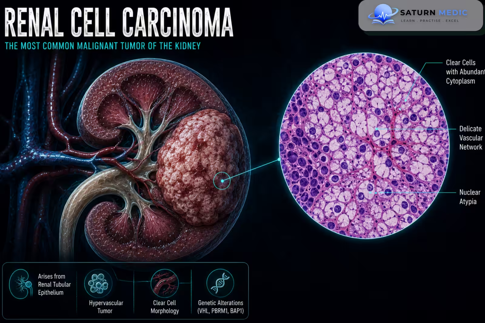
Renal Cell Carcinoma (RCC) is a malignant tumor arising from the epithelial cells of the renal tubules and is the most common type of kidney cancer in adults. The most common subtype is clear cell RCC.
Also called
ICD-10
Specialty
Onset
Reviewed
-
OverviewOverview
-
Etiology & Risk FactorsEtiology & Risk Factors
-
PathophysiologyPathophysiology
-
Clinical PresentationClinical Presentation
-
History TakingHistory Taking
-
Physical ExaminationPhysical Examination
-
InvestigationsInvestigations
-
DiagnosisDiagnosis
-
ManagementManagement
-
ComplicationsComplications
-
PrognosisPrognosis
-
Key Points / Clinical PearlsKey Points / Clinical Pearls
Overview
–Renal Cell Carcinoma (RCC) includes several histological subtypes. The main types include:
- Clear cell RCC — most common
- Papillary RCC
- Chromophobe RCC
The prognosis and management of Renal Cell Carcinoma (RCC) depend primarily on the tumor stage, histological subtype, grade, local invasion, and presence of metastases.
Etiology & Risk Factors
-Etiology
– The exact cause of Renal Cell Carcinoma (RCC) is not fully understood. The disease develops through genetic and molecular abnormalities that result in uncontrolled proliferation of renal tubular epithelial cells.
Clear cell RCC is strongly associated with abnormalities involving the von Hippel-Lindau (VHL) pathway.
-Risk Factors
- Increasing age
- Cigarette smoking
- Obesity
- Hypertension
- Chronic Kidney Disease (CKD)
- Long-term dialysis
- Family history of kidney cancer
- Certain hereditary cancer syndromes
Pathophysiology
Genetic and molecular abnormalities → uncontrolled proliferation of renal tubular epithelial cells → tumor formation → progressive local growth → invasion of the renal vein or surrounding tissues → lymphatic and hematogenous spread → metastatic disease
Clear cell RCC commonly involves abnormalities in the VHL pathway, which can promote angiogenesis and tumor growth.
Clinical Presentation
-Symptoms:
Early Renal Cell Carcinoma (RCC) is often asymptomatic.
Possible symptoms include:
- Painless hematuria
- Flank pain
- Abdominal or flank mass
- Fatigue
- Fever
- Unintentional weight loss
- Reduced appetite
-Signs:
- Palpable flank or abdominal mass
- Persistent or newly developed varicocele
- Lower-limb edema
- Hypertension
- Signs of metastatic disease
History Taking
-Ask about:
- Hematuria
- Flank or abdominal pain
- Feeling a mass
- Unintentional weight loss
- Fever or fatigue
- Smoking history
- Hypertension
- Chronic Kidney Disease (CKD)
- Previous kidney tumors
Physical Examination
-General Examination
- Assess general condition
- Look for weight loss or fever
- Measure blood pressure
- Assess for peripheral edema
-System-Specific Examination:
- Palpate the abdomen and flanks for a mass
- Assess for varicocele
- Examine for signs of metastatic disease when indicated
Investigations
Complete Blood Count
Useful because Renal Cell Carcinoma (RCC) may be associated with:
Anemia
Polycythemia in some patients
-Biochemistry / Specific Tests
Serum creatinine
Estimated Glomerular Filtration Rate (eGFR)
Liver function tests
Serum calcium when indicated
There is no specific blood tumor marker for Renal Cell Carcinoma (RCC).
-Imaging
Contrast-Enhanced Computed Tomography (CT)
The main imaging investigation for:
Characterizing a renal mass
Assessing tumor size
Detecting local invasion
Assessing renal vein involvement
Staging the disease
Magnetic Resonance Imaging (MRI)
Useful when CT contrast cannot be used or when detailed assessment of venous involvement is required.
-Special / Confirmatory Tests
Renal Mass Biopsy
Biopsy may be performed when histological confirmation will change management, such as before systemic therapy or selected ablative treatments.
A biopsy is not required before surgery for every patient with a typical resectable renal mass.
Diagnosis
-Renal Cell Carcinoma (RCC) is assessed through:
Suspicious symptoms or incidental renal mass → Contrast-enhanced CT or MRI → Characterization and staging → Renal mass biopsy when required → Definitive treatment and histological confirmation.
Renal Cell Carcinoma · TNM Classification & Staging
| Category | T / N / M | Definition |
|---|---|---|
| Tumour (T) | T1a | Tumour ≤4 cm, confined to kidney |
| T1b | Tumour >4 cm but ≤7 cm, confined to kidney | |
| T2a | Tumour >7 cm but ≤10 cm, confined to kidney | |
| T2b | Tumour >10 cm, confined to kidney | |
| Advanced (T) | T3a | Renal vein / segmental branches, perinephric fat, or renal sinus fat invasion (not beyond Gerota's fascia) |
| T3b | IVC below diaphragm | |
| T3c | IVC above diaphragm or IVC wall invasion | |
| T4 | Beyond Gerota's fascia or direct invasion of adrenal gland | |
| Nodes (N) | N0 N1 | N0 = no regional nodes | N1 = regional lymph node metastasis |
| Metastasis (M) | M0 M1 | M0 = no distant metastasis | M1 = distant metastasis present |
- Tumour ≤7 cm, kidney-confined
- 5-yr survival ~93%
- Surgery: partial or radical nephrectomy
- Active surveillance for small T1a (<3 cm)
- Tumour >7 cm, still kidney-confined
- 5-yr survival ~75%
- Radical nephrectomy standard
- No adjuvant therapy routinely
- Perinephric / vascular extension or nodal disease
- 5-yr survival ~53%
- Radical nephrectomy + thrombus removal if IVC involved
- Adjuvant pembrolizumab (high-risk) — 1 year
- Beyond Gerota's fascia or distant metastasis
- 5-yr survival ~8%
- Systemic therapy: IO + TKI (nivolumab + cabozantinib)
- Cytoreductive nephrectomy selected cases
Renal Cell Carcinoma · WHO/ISUP Grading & Histological Types
| Subtype | Frequency | Key features | Mutation | Prognosis |
|---|---|---|---|---|
| Clear Cell RCC | 70–80% | Clear cytoplasm (glycogen/lipid). Highly vascular. Most common metastatic type. | VHL loss (chr 3p) | Intermediate |
| Papillary RCC | 10–15% | Type 1 (low grade, MET) — indolent. Type 2 (high grade, SETD2/FH) — aggressive. | MET (Type 1), FH (Type 2) | Favourable (Type 1) |
| Chromophobe RCC | 5% | Pale cytoplasm, perinuclear halo. Arises from collecting duct intercalated cells. | Multiple chr losses | Favourable |
| Collecting Duct RCC | <1% | Medullary origin. Highly aggressive. Often metastatic at presentation. | NF2, SETD2 | Poor |
| Medullary RCC | Rare | Sickle cell trait/disease. Young patients. Extremely aggressive. | SMARCB1 loss | Very poor |
Management
1. Definitive Treatment
Localized Disease
Surgery is the main curative treatment.
Options include:
Partial nephrectomy when technically feasible
Radical nephrectomy for larger or more complex tumors
Selected small tumors may be managed with active surveillance or local ablative treatment.
2. Medical Treatment
Advanced Renal Cell Carcinoma (RCC) may be treated with:
Immune checkpoint inhibitor-based therapy
Targeted therapy
Combination systemic therapy
Treatment depends on the subtype, stage, risk category, and patient factors.
3. Surgical / Procedural Treatment
Partial nephrectomy
Radical nephrectomy
Thermal ablation in selected patients
Selected metastasectomy
4. Supportive Management
Monitor renal function
Control blood pressure
Smoking cessation
Pain management
Bone-directed treatment when indicated
Complications
- Local tumor invasion
- Severe hematuria
- Renal vein invasion
- Inferior vena cava tumor thrombus
- Metastatic disease
- Bone metastases
- Pathological fractures
- Brain metastases
Prognosis
The prognosis of Renal Cell Carcinoma (RCC) depends mainly on tumor stage, grade, subtype, tumor size, venous invasion, lymph node involvement, and metastatic spread. Localized disease generally has a favorable prognosis after curative treatment, while metastatic disease has a less favorable prognosis.
Key Points / Clinical Pearls
- Renal Cell Carcinoma (RCC) is the most common kidney cancer in adults.
- Clear cell RCC is the most common subtype.
- Many cases are found incidentally on imaging.
- Early RCC is often asymptomatic.
- Hematuria, flank pain, and a palpable mass are the classic but uncommon triad.
- Smoking, obesity, and hypertension are important risk factors.
- VHL pathway abnormalities are important in clear cell RCC.
- RCC can invade the renal vein and inferior vena cava.
- A persistent new varicocele can suggest an underlying renal mass.
- Contrast-enhanced CT is central to diagnosis and staging.
- There is no specific diagnostic blood test for RCC.
- Biopsy is performed selectively.
- European Association of Urology (EAU). EAU Guidelines on Renal Cell Carcinoma.
- American Urological Association (AUA). Renal Mass and Localized Renal Cancer Guideline.
- National Comprehensive Cancer Network (NCCN). NCCN Guidelines for Kidney Cancer.
- Ljungberg B, et al. European Association of Urology Guidelines on Renal Cell Carcinoma. PubMed.
- National Cancer Institute (NCI). Kidney Cancer.