

Clinical Subject Page
Staghorn Calculus
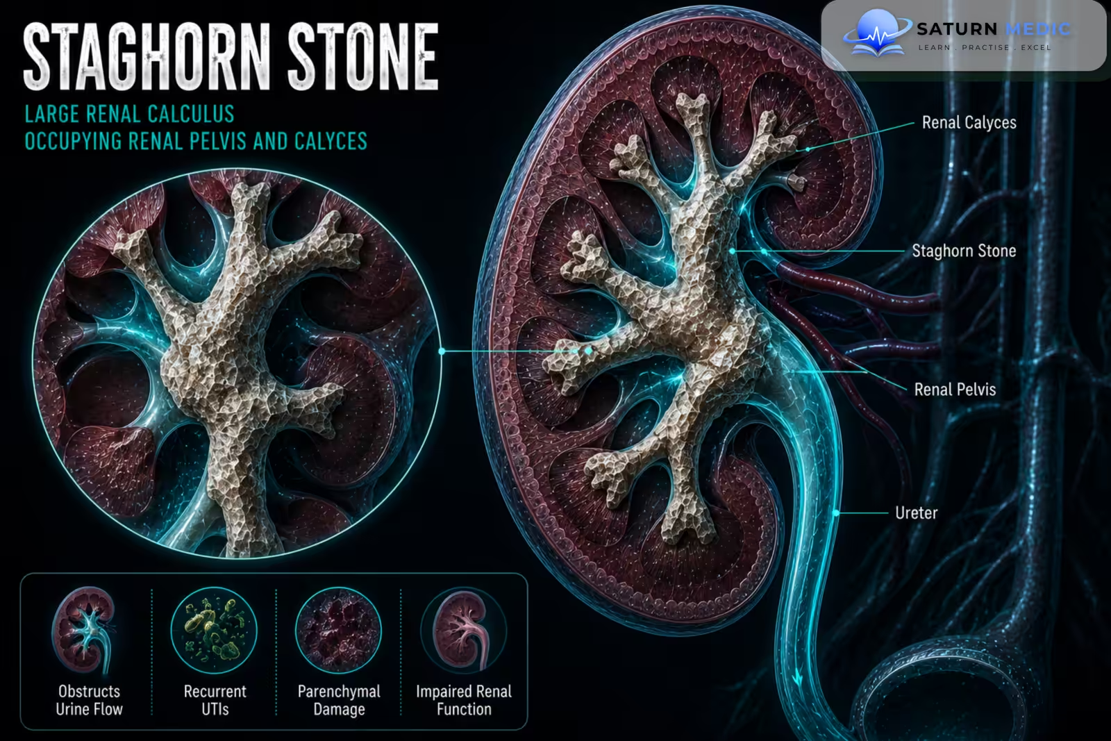
A Staghorn Calculus is a large, branching kidney stone that occupies the renal pelvis and extends into one or more renal calyces, resembling the antlers of a stag. It can cause recurrent infection, obstruction, and progressive kidney damage
Also called
ICD-10
Specialty
Onset
Reviewed
-
OverviewOverview
-
Etiology & Risk FactorsEtiology & Risk Factors
-
PathophysiologyPathophysiology
-
Clinical PresentationClinical Presentation
-
History TakingHistory Taking
-
Physical ExaminationPhysical Examination
-
InvestigationsInvestigations
-
DiagnosisDiagnosis
-
ManagementManagement
-
ComplicationsComplications
-
PrognosisPrognosis
-
Key Points / Clinical PearlsKey Points / Clinical Pearls
Overview
–A Staghorn Calculus is classified according to its extent:
- Partial staghorn calculus: occupies the renal pelvis and extends into one or more calyces.
- Complete staghorn calculus: occupies the renal pelvis and extends into most or all major calyces.
–Unlike a small urinary stone, a Staghorn Calculus may cause relatively mild symptoms despite extensive stone burden. Without treatment, it can lead to recurrent infection, renal scarring, loss of kidney function, and Chronic Kidney Disease CKD.
Etiology & Risk Factors
-Etiology
–Classic infection-related Staghorn Calculus develops due to infection with urease-producing bacteria. These organisms produce urease, which increases urinary ammonia and raises urine pH.
This promotes formation of magnesium ammonium phosphate, also called struvite.
Common urease-producing organisms include:
- Proteus species
- Klebsiella species
- Other urease-producing bacteria
Not all staghorn calculi are struvite stones.
-Risk Factors
- Recurrent Urinary Tract Infection (UTI)
- Infection with urease-producing organisms
- Urinary tract obstruction
- Neurogenic bladder
- Urinary stasis
Pathophysiology
Urease-producing bacterial infection → urea breakdown → increased ammonia production → alkaline urine → struvite crystal formation → progressive stone growth → branching occupation of renal pelvis and calyces → recurrent infection and obstruction → renal scarring and loss of kidney function
Clinical Presentation
-Symptoms:
- Dull or persistent flank pain
- Recurrent Urinary Tract Infection (UTI)
- Fever
- Dysuria
- Hematuria
- Malaise
- Passage of stone fragments
-Signs:
- Costovertebral angle tenderness
- Fever during active infection
- Tachycardia or hypotension in severe infection
- Signs of sepsis
History Taking
-Ask about:
- Flank pain
- Recurrent UTI
- Fever or rigors
- Dysuria
- Hematuria
- Previous urinary stones
- Previous stone analysis
- Previous urological procedures
- Urinary tract obstruction
- Neurogenic bladder
- Previous kidney infections
Physical Examination
-General Examination
- Record temperature and vital signs
- Assess for systemic infection or sepsis
- Assess hydration status
-System-Specific Examination:
- Check for costovertebral angle tenderness
- Examine the abdomen for tenderness
- Assess for signs of urinary obstruction when relevant
Investigations
Biochemistry / Specific Tests
Urinalysis
Urine culture and sensitivity
Serum creatinine
Estimated Glomerular Filtration Rate (eGFR)
Serum electrolytes when kidney dysfunction is present
Stone analysis after removal
Metabolic evaluation may be required in selected patients.
-Imaging
Non-contrast Computed Tomography (CT)
The main imaging investigation for assessing:
Stone size
Branching extent
Renal anatomy
Stone density
Treatment planning
Plain X-ray of the Kidneys, Ureters, and Bladder (KUB)
May be useful for radiopaque stones and follow-up.
-Special / Confirmatory Tests
Renal functional assessment may be required when there is concern about significant loss of function in the affected kidney.
Diagnosis
-Staghorn Calculus is diagnosed by:
Clinical suspicion or recurrent UTI → urinalysis and urine culture → assessment of renal function → CT imaging demonstrating a branching stone occupying the renal pelvis and extending into the calyces.
CT is also essential for planning definitive treatment.
Management
1. First-Line / Emergency Management
There is no routine emergency treatment for uncomplicated disease.
If sepsis or an infected obstructed kidney is present:
Start appropriate antibiotics
Urgently drain the collecting system when indicated
Stabilize the patient
Definitive stone treatment should be delayed until infection is adequately controlled.
2. Definitive Treatment
The main goal is complete stone clearance whenever safely possible.
Percutaneous Nephrolithotomy (PCNL) is the preferred definitive treatment for most large or complex staghorn stones.
3. Medical Treatment
Culture-directed antibiotics for proven infection
Analgesics when required
Metabolic treatment based on stone composition and risk factors
4. Surgical / Procedural Treatment
Percutaneous Nephrolithotomy (PCNL)
Staged PCNL for complex stones
Flexible ureteroscopy as an adjunct in selected patients
Shock Wave Lithotripsy (SWL) for selected residual fragments
Nephrectomy when the affected kidney is non-functioning and removal is clinically appropriate
5. Supportive Management
Monitor renal function
Treat recurrent UTI
Maintain appropriate hydration
Complications
- Recurrent Urinary Tract Infection (UTI)
- Pyelonephritis
- Urosepsis
- Hydronephrosis
- Renal scarring
- Acute Kidney Injury (AKI)
- Chronic Kidney Disease CKD
- Loss of renal function
- Stone recurrence
Prognosis
The prognosis of Staghorn Calculus depends on the degree of infection, baseline renal function, completeness of stone removal, and prevention of recurrence. Early treatment can preserve kidney function, while untreated disease may result in recurrent infection and irreversible renal damage.
Key Points / Clinical Pearls
- Staghorn Calculus is a large branching kidney stone.
- It occupies the renal pelvis and extends into the renal calyces.
- It may be partial or complete.
- Classic infection-related stones are often composed of struvite.
- Struvite formation is associated with urease-producing bacteria.
- Proteus species are an important associated organism.
- Symptoms may be mild despite extensive stone burden.
- Recurrent UTI is a common presentation.
- Untreated disease can cause progressive kidney damage.
- Urine culture is important before intervention.
- CT is essential for defining stone anatomy and planning treatment.
- Percutaneous Nephrolithotomy (PCNL) is the main treatment for most cases.
- European Association of Urology (EAU). EAU Guidelines on Urolithiasis .
- American Urological Association (AUA). Surgical Management of Stones Guideline .
- Assimos D, Krambeck A, Miller NL, et al. Surgical Management of Stones: American Urological Association/Endourological Society Guideline. J Urol. 2016;196(4):1153-1160. Journal of Urology .
- Diri A, Diri B. Management of Staghorn Renal Stones. Ren Fail. 2018;40(1):357-362. PubMed .
- Torricelli FCM, Monga M. Staghorn Renal Stones: What the Urologist Needs to Know. Curr Opin Urol. 2015;25(4):326-331. PubMed .
- National Library of Medicine (NIH). Staghorn Renal Stones . StatPearls.