

Clinical Subject Page
Testicular Cancer
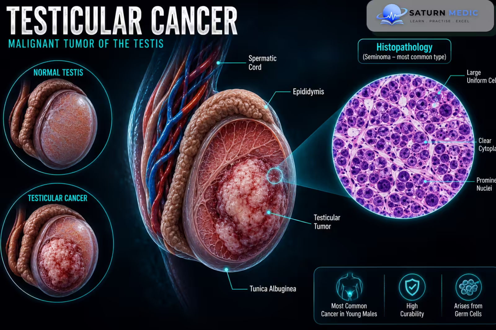
Testicular Cancer is a malignant tumor arising from the testis and is the most common solid malignancy in young adult males. Most Testicular Cancer cases are germ cell tumors, which are broadly classified into seminomas and non-seminomatous germ cell tumors
Also called
ICD-10
Specialty
Onset
Reviewed
-
OverviewOverview
-
Etiology & Risk FactorsEtiology & Risk Factors
-
PathophysiologyPathophysiology
-
Clinical PresentationClinical Presentation
-
History TakingHistory Taking
-
Physical ExaminationPhysical Examination
-
InvestigationsInvestigations
-
DiagnosisDiagnosis
-
ManagementManagement
-
ComplicationsComplications
-
PrognosisPrognosis
-
Key Points / Clinical PearlsKey Points / Clinical Pearls
Overview
–Most Testicular Cancer occurs in younger males and arises from germ cells. The two major groups are:
- Seminoma: usually more radiosensitive and tends to spread more predictably.
- Non-seminomatous germ cell tumor (NSGCT): includes embryonal carcinoma, yolk sac tumor, choriocarcinoma, and teratoma.
Mixed germ cell tumors are also common.
Testicular Cancer often spreads first to the retroperitoneal lymph nodes and may later metastasize to the lungs, liver, brain, or other organs.
Etiology & Risk Factors
-Etiology
–The exact cause of Testicular Cancer is not fully understood. Most tumors develop from abnormalgerm cells within the testis and are associated with characteristic genetic and developmental abnormalities.
-Risk Factors
- Cryptorchidism
- Previous Testicular Cancer
- Family history
- Testicular dysgenesis or impaired testicular development
- Infertility
Pathophysiology
Abnormal germ cell development → accumulation of genetic abnormalities → germ cell neoplasia → invasive Testicular Cancer → local tumor growth → lymphatic spread to retroperitoneal lymph nodes → distant metastasis
Clinical Presentation
-Symptoms:
Testicular Cancer commonly presents with:
- Painless testicular lump
- Unilateral testicular enlargement
- Testicular heaviness
- Dull testicular or scrotal discomfort
Less commonly:
- Acute pain due to hemorrhage or infarction within the tumor
- Gynecomastia
- Back pain from retroperitoneal lymphadenopathy
- Cough or dyspnea from pulmonary metastases
-Signs:
- Firm intratesticular mass
- Enlarged testis
- Irregular testicular contour
- Gynecomastia in selected hormone-producing tumors
History Taking
-Ask about:
- Duration of the testicular lump
- Pain or discomfort
- Testicular enlargement
- Feeling of heaviness
- Previous Cryptorchidism
- Previous Testicular Cancer
- Family history
- Fertility history
- Gynecomastia
- Back or abdominal pain
Physical Examination
-General Examination
- Assess general condition
- Examine for gynecomastia
- Look for signs of metastatic disease
-System-Specific Examination:
- Assess general condition
- Examine for gynecomastia
- Look for signs of metastatic disease
Investigations
-Biochemistry / Specific Tests
Serum Tumor Markers
Measure:
Alpha-fetoprotein (AFP)
Beta-human Chorionic Gonadotropin (β-hCG)
Lactate Dehydrogenase (LDH)
AFP should not be elevated in pure seminoma. Tumor markers are useful for diagnosis, staging, risk assessment, and monitoring.
-Imaging
Scrotal Ultrasound
The first-line imaging investigation for a suspected testicular mass.
Computed Tomography (CT)
Used for staging, particularly assessment of retroperitoneal lymph nodes and distant metastases.
-Special / Confirmatory Tests
Radical Inguinal Orchiectomy
Provides definitive histological diagnosis and is the standard initial treatment for most suspected testicular malignancies.
Diagnosis
-Testicular Cancer is diagnosed through:
Painless testicular mass → scrotal ultrasound → serum tumor markers → radical inguinal orchiectomy for definitive histology → staging imaging and post-orchiectomy tumor marker assessment.
Management
Testicular Cancer · Seminoma Management by Stage
| Stage | Definition | Treatment Options | Cure Rate |
|---|---|---|---|
| Stage I | Confined to testis, no metastasis |
1st Active surveillance — preferred; 15–20% relapse but salvageable Alt Adjuvant carboplatin ×1 cycle (AUC 7) — ↓ relapse to <5% Rarely Para-aortic RT (20 Gy) — if surveillance not feasible |
~99% |
| Stage IIA/B | Retroperitoneal nodes ≤5 cm |
Standard Para-aortic + iliac RT (30–36 Gy) Alt BEP ×3 cycles (bleomycin, etoposide, cisplatin) — IIB preferred |
~95% |
| Stage IIC / III | Nodes >5 cm or distant metastasis |
Standard BEP ×3–4 cycles (good prognosis) or EP ×4 cycles Intermediate/poor: BEP ×4 cycles Residual mass >3 cm post-chemo → PET-CT; if PET+ → salvage |
~90% |
Testicular Cancer · Non-Seminoma (NSGCT) Management by Stage
| Stage | Definition | Treatment Options | Cure Rate |
|---|---|---|---|
| Stage I | Confined to testis; markers normalise post-orchidectomy |
Low risk Active surveillance — no LVI, no embryonal CA predominance High risk BEP ×1 cycle — LVI present; ↓ relapse from 50% → <5% Alt Nerve-sparing RPLND (retroperitoneal lymph node dissection) |
~99% |
| Stage IIA/B | Retroperitoneal nodes, markers normal or mildly raised |
Standard BEP ×3 cycles (good prognosis) or primary RPLND (IIA, markers −) Post-chemo residual mass → RPLND (teratoma cannot be killed by chemo) |
~95% |
| Stage III (Good) | Non-pulmonary mets absent; markers: AFP <1000, hCG <5000, LDH <1.5×ULN |
Standard BEP ×3 cycles Post-chemo residual mass → RPLND mandatory |
~95% |
| Stage III (Intermediate / Poor) | Non-pulmonary visceral mets OR very high markers |
Standard BEP ×4 cycles Poor prognosis: consider VIP or high-dose chemo + ASCT Post-chemo residual → RPLND |
~50–70% |
Complications
- Retroperitoneal lymph node metastases
- Pulmonary metastases
- Liver metastases
- Brain metastases
- Infertility
- Hypogonadism
- Treatment-related cardiovascular complications
- Secondary malignancy
- Psychological distress
Prognosis
The prognosis of Testicular Cancer is generally excellent. It is one of the most curable solid cancers, including many cases with metastatic disease. Prognosis depends on histological subtype, stage, tumor marker levels, metastatic sites, and response to treatment.
Key Points / Clinical Pearls
- Testicular Cancer is most common in young adult males.
- Most cases are germ cell tumors.
- Seminoma and NSGCT are the major groups.
- A painless testicular mass is the classic presentation.
- Cryptorchidism is a major risk factor.
- Scrotal ultrasound is the first-line investigation.
- AFP, β-hCG, and LDH are important tumor markers.
- AFP elevation excludes pure seminoma.
- Normal tumor markers do not exclude Testicular Cancer.
- Radical inguinal orchiectomy provides diagnosis and initial treatment.
- Transscrotal biopsy should be avoided.
- Retroperitoneal lymph nodes are a common initial site of spread.
- European Association of Urology (EAU). EAU Guidelines on Testicular Cancer.
- American Urological Association (AUA). Diagnosis and Treatment of Early-Stage Testicular Cancer.
- National Comprehensive Cancer Network (NCCN). NCCN Guidelines for Testicular Cancer.
- Gilligan T, Lin DW, Aggarwal R, et al. Testicular Cancer, Version 2.2020, NCCN Clinical Practice Guidelines in Oncology. PubMed.
- National Cancer Institute (NCI). Testicular Cancer.