

Clinical Subject Page
Ureteral Stones
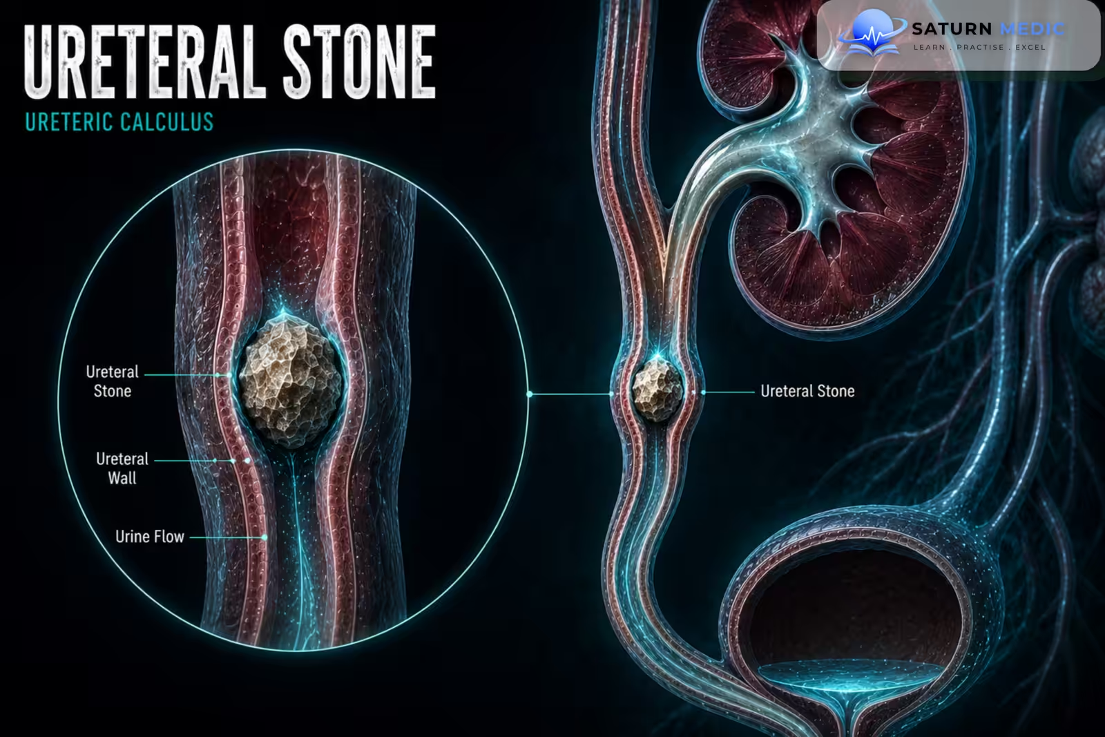
Ureteral Stones are urinary calculi located within one or both ureters. Most Ureteral Stones originate in the kidneys and migrate into the ureter. They can obstruct urine flow and typically cause sudden, severe colicky flank pain known as renal colic
Also called
ICD-10
Specialty
Onset
Reviewed
-
OverviewOverview
-
Etiology & Risk FactorsEtiology & Risk Factors
-
PathophysiologyPathophysiology
-
Clinical PresentationClinical Presentation
-
History TakingHistory Taking
-
Physical ExaminationPhysical Examination
-
InvestigationsInvestigations
-
DiagnosisDiagnosis
-
ManagementManagement
-
ComplicationsComplications
-
PrognosisPrognosis
-
Key Points / Clinical PearlsKey Points / Clinical Pearls
Overview
–The clinical importance of Ureteral Stones depends on their size, location, degree of obstruction, and the presence of infection.
Stones commonly become lodged at three anatomical narrowing points:
- Ureteropelvic Junction (UPJ)
- Crossing of the iliac vessels
- Ureterovesical Junction (UVJ)
-An infectedobstructed urinarysystem caused by Ureteral Stones is a urological emergency requiring urgent drainage.
Etiology & Risk Factors
-Etiology
–Most Ureteral Stonesform in the kidney before migrating into the ureter. Common stone compositions include:
- Calcium oxalate
- Calcium phosphate
- Uric acid
- Struvite
- Cystine
-Risk Factors
- Previous urinary stones
- Low fluid intake
- Metabolic abnormalities
- Recurrent Urinary Tract Infection (UTI)
- Family history of stone disease
Pathophysiology
Urinary supersaturation → crystal formation in the kidney → stone growth → migration into the ureter → ureteral obstruction and spasm → increased upstream pressure → ureteral dilation → renal colic and possible Hydronephrosis
Clinical Presentation
-Symptoms:
- Sudden severe unilateral flank pain
- Colicky pain occurring in waves
- Pain radiating to the lower abdomen, groin, or genitalia
- Nausea and vomiting
- Hematuria
- Dysuria with distal stones
-Signs:
- Severe discomfort and restlessness
- Costovertebral angle tenderness
- Abdominal tenderness
- Tachycardia due to pain
History Taking
-Ask about:
- Onset and severity of pain
- Radiation to the groin or genitalia
- Previous urinary stones
- Hematuria
- Dysuria, frequency, or urgency
- Fever or rigors
- Nausea and vomiting
- Reduced urine output
- Fluid intake
- Family history of stones
Physical Examination
-General Examination
- Assess pain severity
- Record temperature and vital signs
- Look for signs of sepsis
- Assess hydration status
-System-Specific Examination:
- Check for costovertebral angle tenderness
- Examine for abdominal tenderness
- Assess for a distended bladder when indicated
Investigations
-Biochemistry / Specific Tests
-
Urinalysis
-
Urine culture when infection is suspected
-
Serum creatinine to assess kidney function
-
Serum electrolytes when AKI or significant obstruction is suspected
Selected patients with recurrent stones may require metabolic evaluation.
-Imaging
Non-contrast Computed Tomography (CT)
Highly accurate for confirming Ureteral Stones, identifying stone size and location, and assessing obstruction.
Ultrasound
Useful for detecting Hydronephrosis and preferred when radiation exposure should be minimized.
-Special / Confirmatory Tests
Stone analysis should be performed when a stone is passed or removed and is available for testing.
Diagnosis
-Ureteral Stones are diagnosed by:
Typical renal colic → urinalysis → assessment for infection and renal impairment → imaging to confirm stone size, location, and obstruction.
-Non-contrast CT is commonly used for definitive diagnosis.
Management
1. First-Line / Emergency Management
Initial management includes:
-
Nonsteroidal anti-inflammatory drugs (NSAIDs) when appropriate
-
Antiemetics for nausea and vomiting
-
Oral or intravenous fluids according to hydration status
Urgent drainage is required for:
-
Obstruction with infection or sepsis
-
Anuria
-
Significant Acute Kidney Injury (AKI)
-
Obstruction in a solitary kidney
2. Definitive Treatment
Small uncomplicated Ureteral Stones may be managed conservatively while awaiting spontaneous passage.
Persistent, large, obstructing, or complicated stones usually require intervention.
3. Medical Treatment
-
NSAIDs for pain
-
Antiemetics
-
Alpha-blockers as medical expulsive therapy in selected patients
Antibiotics are indicated only for proven infection.
4. Surgical / Procedural Treatment
-
Ureteroscopy with stone extraction or fragmentation
-
Shock Wave Lithotripsy (SWL)
-
Ureteric stent placement
-
Percutaneous nephrostomy for urgent drainage
5. Supportive Management
-
Maintain appropriate hydration
-
Strain urine when advised
-
Analyze retrieved stones
-
Assess recurrence risk
Complications
- Hydronephrosis
- Urinary Tract Infection (UTI)
- Pyelonephritis
- Urosepsis
- Acute Kidney Injury (AKI)
- Persistent obstruction
- Ureteral stricture
- Recurrent stones
Prognosis
The prognosis of Ureteral Stones is generally excellent when obstruction and infection are treated promptly. Small stones frequently pass spontaneously, while larger stones may require intervention. Outcome depends on stone size, location, infection, duration of obstruction, renal function, and recurrence risk.
Key Points / Clinical Pearls
- Nephrolithiasis means stone formation within the kidneys or urinary tract.
- Calcium oxalate is the most common type of urinary stone.
- Low urine volume is a major modifiable risk factor.
- Severe colicky flank pain radiating to the groin is a classic presentation.
- Hematuria commonly occurs.
- Fever with an obstructing stone suggests a potentially life-threatening infection.
- Urinalysis is an important initial investigation.
- Non-contrast CT KUB is the most accurate imaging test for most adults.
- Ultrasound is useful when radiation should be avoided.
- NSAIDs are usually first-line treatment for acute renal colic when appropriate.
- Small stones may pass spontaneously.
- Larger or obstructing stones may require ureteroscopy, ESWL, or PCNL.
- European Association of Urology (EAU). EAU Guidelines on Urolithiasis .
- American Urological Association (AUA). Surgical Management of Stones Guideline .
- Assimos D, Krambeck A, Miller NL, et al. Surgical Management of Stones: American Urological Association/Endourological Society Guideline. J Urol. 2016;196(4):1153-1160. Journal of Urology .
- Türk C, Neisius A, Petrik A, et al. EAU Guidelines on Urolithiasis. European Association of Urology. Full Guideline .
- Pearle MS, Goldfarb DS, Assimos DG, et al. Medical Management of Kidney Stones: AUA Guideline. J Urol. 2014;192(2):316-324. Journal of Urology .
- National Library of Medicine (NIH). Ureterolithiasis . StatPearls.