

Clinical Subject Page
Upper Tract Urothelial Carcinoma (UTUC)
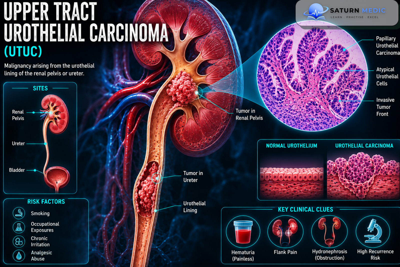
Upper Tract Urothelial Carcinoma (UTUC) is a malignant tumor arising from the urothelial lining of the upper urinary tract, including the renal pelvis and ureter. It is less common than bladder cancer but can be aggressive, particularly when invasive.
Also called
ICD-10
Specialty
Onset
Reviewed
-
OverviewOverview
-
Etiology & Risk FactorsEtiology & Risk Factors
-
PathophysiologyPathophysiology
-
Clinical PresentationClinical Presentation
-
History TakingHistory Taking
-
Physical ExaminationPhysical Examination
-
InvestigationsInvestigations
-
DiagnosisDiagnosis
-
ManagementManagement
-
ComplicationsComplications
-
PrognosisPrognosis
-
Key Points / Clinical PearlsKey Points / Clinical Pearls
Overview
–Upper Tract Urothelial Carcinoma (UTUC) develops from urothelial cells lining the renal pelvis and ureter. It is broadly assessed according to tumor grade, depth of invasion, location, and metastatic spread.
Clinically, UTUC may be categorized as:
- Low-risk disease: more likely to be localized and suitable for kidney-sparing treatment in selected patients.
- High-risk disease: associated with invasive disease and often requires radical treatment.
Etiology & Risk Factors
-Etiology
–Upper Tract Urothelial Carcinoma (UTUC) develops through genetic and molecular abnormalities within urothelial cells. Prolonged exposure of the urothelium to carcinogens contributes to malignant transformation.
-Risk Factors
- Cigarette smoking
- Occupational exposure to certain chemicals
- Increasing age
- Previous or concurrent bladder cancer
- Lynch syndrome
- Exposure to aristolochic acid
Pathophysiology
Exposure to carcinogens and genetic abnormalities → urothelial DNA damage → accumulation of mutations → uncontrolled urothelial cell proliferation → tumor formation in the renal pelvis or ureter → local invasion → lymphatic and hematogenous spread
Clinical Presentation
-Symptoms:
- Visible or microscopic hematuria
- Flank pain
- Flank mass, rarely
- Dysuria
- Urinary frequency
- Recurrent Urinary Tract Infection (UTI)
Flank pain may occur when the tumor obstructs the ureter and causes hydronephrosis.
-Signs:
Early disease may have no specific physical findings.
Advanced disease may cause:
- Flank mass
- Weight loss
- Peripheral edema
- Lymphadenopathy
History Taking
-Ask about:
- Visible or microscopic hematuria
- Flank pain
- Previous bladder cancer
- Dysuria or lower urinary tract symptoms
- Smoking history
- Occupational chemical exposure
- Family history of Lynch syndrome
- Previous urinary tract tumors
- Recurrent UTI
- Weight loss
Physical Examination
-General Examination
- Assess general condition
- Look for weight loss
- Check for peripheral edema
- Assess for lymphadenopathy when advanced disease is suspected
-System-Specific Examination:
- Examine the abdomen and flanks for masses
- Assess for flank tenderness
- Look for signs of urinary obstruction
Investigations
-Biochemistry / Specific Tests
Urinalysis
Urine culture when infection is suspected
Serum creatinine and estimated Glomerular Filtration Rate (eGFR)
Urine cytology, particularly for suspected high-grade disease
-Imaging
Computed Tomography Urography (CT Urography)
The main imaging investigation for suspected Upper Tract Urothelial Carcinoma (UTUC).
It can assess:
Tumor location
Filling defects
Ureteric obstruction
Hydronephrosis
Regional lymph nodes
Other urinary tract abnormalities
-Special / Confirmatory Tests
Diagnostic Ureteroscopy
Allows direct visualization of suspicious lesions.
Ureteroscopic Biopsy
May provide tissue for histological confirmation and tumor grading.
Cystoscopy
Should be performed to evaluate the bladder because synchronous bladder tumors may occur.
Diagnosis
-Upper Tract Urothelial Carcinoma (UTUC) is assessed through:
Hematuria or suspicious symptoms → CT urography → cystoscopy to evaluate the bladder → diagnostic ureteroscopy ± biopsy when required → risk and stage assessment → definitive treatment planning.
Management
1. Definitive Treatment
Treatment depends on risk classification, tumor location, kidney function, and patient factors.
Low-Risk UTUC
Kidney-sparing treatment may be appropriate in selected patients:
Endoscopic tumor ablation
Segmental ureterectomy in selected cases
High-Risk UTUC
Radical nephroureterectomy with removal of a cuff of bladder is the standard treatment for many patients with high-risk localized disease.
2. Medical Treatment
Systemic therapy may be used:
Before surgery in selected high-risk patients
After surgery in appropriate patients
For locally advanced or metastatic disease
Treatment may include platinum-based chemotherapy and other systemic therapies depending on disease characteristics.
3. Surgical / Procedural Treatment
Endoscopic tumor ablation
Segmental ureterectomy
Radical nephroureterectomy
Bladder cuff excision
4. Supportive Management
Monitor renal function
Smoking cessation
Manage urinary obstruction
Treat infection
Regular cystoscopic surveillance
Complications
- Ureteric obstruction
- Hydronephrosis
- Kidney dysfunction
- Local tumor invasion
- Lymph node metastases
- Distant metastases
- Bladder tumor recurrence
- Contralateral upper urinary tract recurrence
Prognosis
The prognosis of Upper Tract Urothelial Carcinoma (UTUC) depends mainly on tumor stage, grade, depth of invasion, lymph node involvement, metastatic spread, and whether complete tumor removal is possible. Low-grade localized disease generally has a better prognosis, while invasive or metastatic disease carries a less favorable outcome.
Key Points / Clinical Pearls
- Upper Tract Urothelial Carcinoma (UTUC) arises in the renal pelvis or ureter.
- Most cases are urothelial carcinomas.
- Hematuria is the most common presentation.
- Flank pain may indicate ureteric obstruction.
- Smoking is an important risk factor.
- Lynch syndrome increases the risk of UTUC.
- Previous bladder cancer is an important clinical risk factor.
- CT urography is the main imaging investigation.
- Cystoscopy is important because bladder tumors may coexist.
- Ureteroscopy can directly visualize the lesion.
- Biopsy helps with diagnosis and risk assessment.
- Selected low-risk tumors can be treated with kidney-sparing approaches.
- Radical nephroureterectomy is standard treatment for many high-risk tumors.
- European Association of Urology (EAU). EAU Guidelines on Upper Urinary Tract Urothelial Carcinoma.
- American Urological Association (AUA). Non-Metastatic Upper Tract Urothelial Carcinoma Guideline.
- National Comprehensive Cancer Network (NCCN). NCCN Guidelines for Urothelial Carcinoma.
- Rouprêt M, Babjuk M, Compérat E, et al. European Association of Urology Guidelines on Upper Urinary Tract Urothelial Carcinoma. PubMed.
- National Cancer Institute (NCI). Urothelial Cancer Information.