
Clinical Subject Page

Hydronephrosis
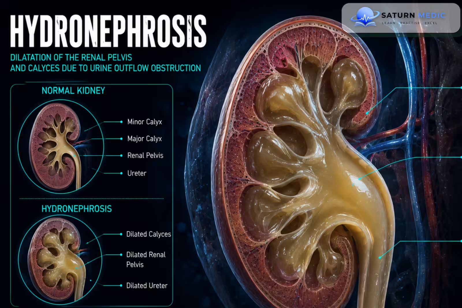
Hydronephrosis is dilatation of the renal pelvis and calyces caused by impaired drainage of urine. Persistent or severe obstruction can increase pressure within the kidney and lead to progressive renal damage
Also called
ICD-10
Specialty
Onset
Reviewed
-
OverviewOverview
-
Etiology & Risk FactorsEtiology & Risk Factors
-
PathophysiologyPathophysiology
-
Clinical PresentationClinical Presentation
-
History TakingHistory Taking
-
Physical ExaminationPhysical Examination
-
InvestigationsInvestigations
-
DiagnosisDiagnosis
-
ManagementManagement
-
ComplicationsComplications
-
PrognosisPrognosis
-
Key Points / Clinical PearlsKey Points / Clinical Pearls
Overview
-Hydronephrosis occurs when urine cannot drain normally from the kidney, causing dilatation of the collecting system.
It may be:
- Unilateral or bilateral
- Acute or chronic
- Mild, moderate, or severe
- Obstructive or non-obstructive
–Common causes include urinary stones, UreteroPelvic Junction obstruction, prostatic enlargement, urethral obstruction, pregnancy, tumors, and congenital abnormalities.
Etiology & Risk Factors
-Etiology:
Hydronephrosis usually results from urinary tract obstruction or impaired urine drainage.
Important causes include:
- Nephrolithiasis causing ureteric obstruction
- Ureteropelvic junction obstruction
- Ureteric stricture
- Benign Prostatic Hyperplasia (BPH)
- Urethral obstruction, including Posterior Urethral Valves (PUV)
- Pelvic or urinary tract tumors
- Pregnancy-related ureteric compression
- Neurogenic bladder
- Vesicoureteral Reflux (VUR)
-Risk Factors
- History of urinary stones
- Congenital urinary tract abnormalities
- Benign Prostatic Hyperplasia (BPH)
- Pelvic malignancy or surgery
- Pregnancy
Pathophysiology
Impaired urinary drainage → urine accumulates proximal to the obstruction → increased pressure in the renal pelvis and calyces → hydronephrosis → compression of renal tissue and reduced renal blood flow → tubular and nephron damage → reduced kidney function → Acute Kidney Injury (AKI) or Chronic Kidney Disease CKD if severe or prolonged
Clinical Presentation
–Symptoms
Hydronephrosis may be asymptomatic, especially when chronic.
Possible symptoms include:
- Flank pain
- Renal colic
- Abdominal discomfort
- Nausea and vomiting
- Difficulty passing urine
- Reduced urine output
- Urinary frequency
- Fever when infection is present
- Hematuria
-Signs:
- Costovertebral angle tenderness
- Flank tenderness
- Palpable kidney in severe cases
- Distended bladder when lower urinary tract obstruction is present
- Fever and systemic illness when associated infection is present
History Taking
-Ask about:
- Flank or abdominal pain
- Sudden or gradual onset of symptoms
- Previous urinary stones
- Hematuria
- Fever or chills
- Dysuria
- Urinary frequency or urgency
- Difficulty voiding
- Poor urinary stream
- Reduced urine output or anuria
- History of Benign Prostatic Hyperplasia (BPH)
Physical Examination
-General Examination
- Temperature
- Blood pressure
- Hydration status
- Signs of sepsis
- Signs of Acute Kidney Injury (AKI) or Chronic Kidney Disease CKD when present
-System-Specific Examination:
- Costovertebral angle tenderness
- Flank or abdominal tenderness
- Palpable abdominal mass in severe hydronephrosis
- Palpable distended bladder in lower urinary tract obstruction
Investigations
-Biochemistry / Specific Tests
Serum creatinine and estimated kidney function
Serum electrolytes when significant obstruction or kidney impairment is suspected
Urinalysis for hematuria, pyuria, and protein
Urine culture when urinary tract infection is suspected
-Imaging
Renal and Bladder Ultrasound
The usual first-line imaging investigation for detecting Hydronephrosis.
It can assess:
Degree of collecting-system dilatation
Kidney size
Bladder distension
Post-void residual urine
Some structural abnormalities
-Non-Contrast CT
Useful when urinary stones are suspected.
-CT Urography or MR Urography
May be used to identify:
Level of obstruction
Ureteric strictures
Tumors
Complex urinary tract abnormalities
-Special / Confirmatory Tests
Further tests depend on the suspected cause and may include:
Diuretic renography to assess functional obstruction
Voiding Cystourethrogram (VCUG) when Vesicoureteral Reflux (VUR) or lower urinary tract obstruction is suspected
Cystoscopy for selected lower urinary tract causes
Diagnosis
-Diagnosis is based on:
Imaging demonstrating dilatation of the renal pelvis and calyces + identification of the underlying cause of impaired urinary drainage.
Renal function should be assessed in significant, bilateral, prolonged, or symptomatic Hydronephrosis
Management
1. First-Line / Emergency Management
Urgent treatment is required when Hydronephrosis is associated with infection, sepsis, anuria, severe Acute Kidney Injury (AKI), or obstruction of a solitary functioning kidney.
Emergency management may include:
Intravenous fluids when appropriate
Analgesia
Antibiotics for infection
Urgent urinary tract decompression
2. Definitive Treatment
Definitive treatment depends on the underlying cause.
Examples include:
Stone treatment or removal
Correction of ureteropelvic junction obstruction
Treatment of ureteric strictures
Management of Benign Prostatic Hyperplasia (BPH)
Treatment of tumors causing obstruction
Correction of congenital abnormalities
3. Medical Treatment
Depending on the cause:
Analgesics
Antibiotics for confirmed or suspected infection
Medical expulsive therapy in selected ureteric stone cases
Medication for Benign Prostatic Hyperplasia (BPH) when appropriate
4. Surgical / Procedural Treatment
Urgent or elective procedures may include:
Ureteric stent placement
Percutaneous nephrostomy
Ureteroscopy
Pyeloplasty
Stone removal procedures
Transurethral or other treatment for bladder outlet obstruction
5. Supportive Management
Monitor urine output
Monitor serum creatinine and electrolytes
Treat the underlying cause
Follow-up imaging
Complications
- Urinary tract infection
- Pyelonephritis
- Urosepsis
- Renal atrophy
- Acute Kidney Injury (AKI)
- Chronic Kidney Disease CKD
- Hypertension
- Permanent loss of renal function
Prognosis
The prognosis depends mainly on the cause, severity, duration of obstruction, presence of infection, and whether one or both kidneys are affected. Acute obstruction relieved promptly may result in substantial recovery of kidney function. Prolonged severe obstruction can cause irreversible renal damage and progressive Chronic Kidney Disease CKD
Key Points / Clinical Pearls
- Hydronephrosis is dilatation of the renal pelvis and calyces due to impaired urine drainage.
- It is an anatomical finding rather than a disease by itself.
- Urinary obstruction is a common cause.
- Nephrolithiasis is an important cause of acute unilateral Hydronephrosis.
- Bilateral Hydronephrosis may indicate lower urinary tract obstruction.
- Renal and bladder ultrasound is the usual first-line investigation.
- CT is particularly useful when urinary stones or another obstructing lesion are suspected.
- Kidney function should be assessed in significant or bilateral disease.
- Hydronephrosis may be asymptomatic when chronic.
- Flank pain, hematuria, and nausea may occur.
- Fever with an obstructed kidney suggests infection and requires urgent treatment.
- An infected obstructed kidney is a urological emergency.
- Urgent decompression may be performed using a ureteric stent or percutaneous nephrostomy.
- European Association of Urology (EAU). EAU Guidelines on Paediatric Urology .
- European Association of Urology (EAU). EAU Guidelines on Urolithiasis .
- American Urological Association (AUA). Kidney Stones: Medical Management Guideline .
- Chevalier RL, Peters CA. Congenital Urinary Tract Obstruction: Proceedings of the State-of-the-Art Strategic Planning Workshop. Pediatr Nephrol. 2003;18(6):576-606. PubMed .
- Nguyen HT, Herndon CDA, Cooper C, et al. The Society for Fetal Urology Consensus Statement on the Evaluation and Management of Antenatal Hydronephrosis. J Pediatr Urol. 2010;6(3):212-231. Journal of Pediatric Urology .
- National Library of Medicine (NIH). Hydronephrosis and Hydroureter . StatPearls.