
Clinical Subject Page

Urethral Stricture
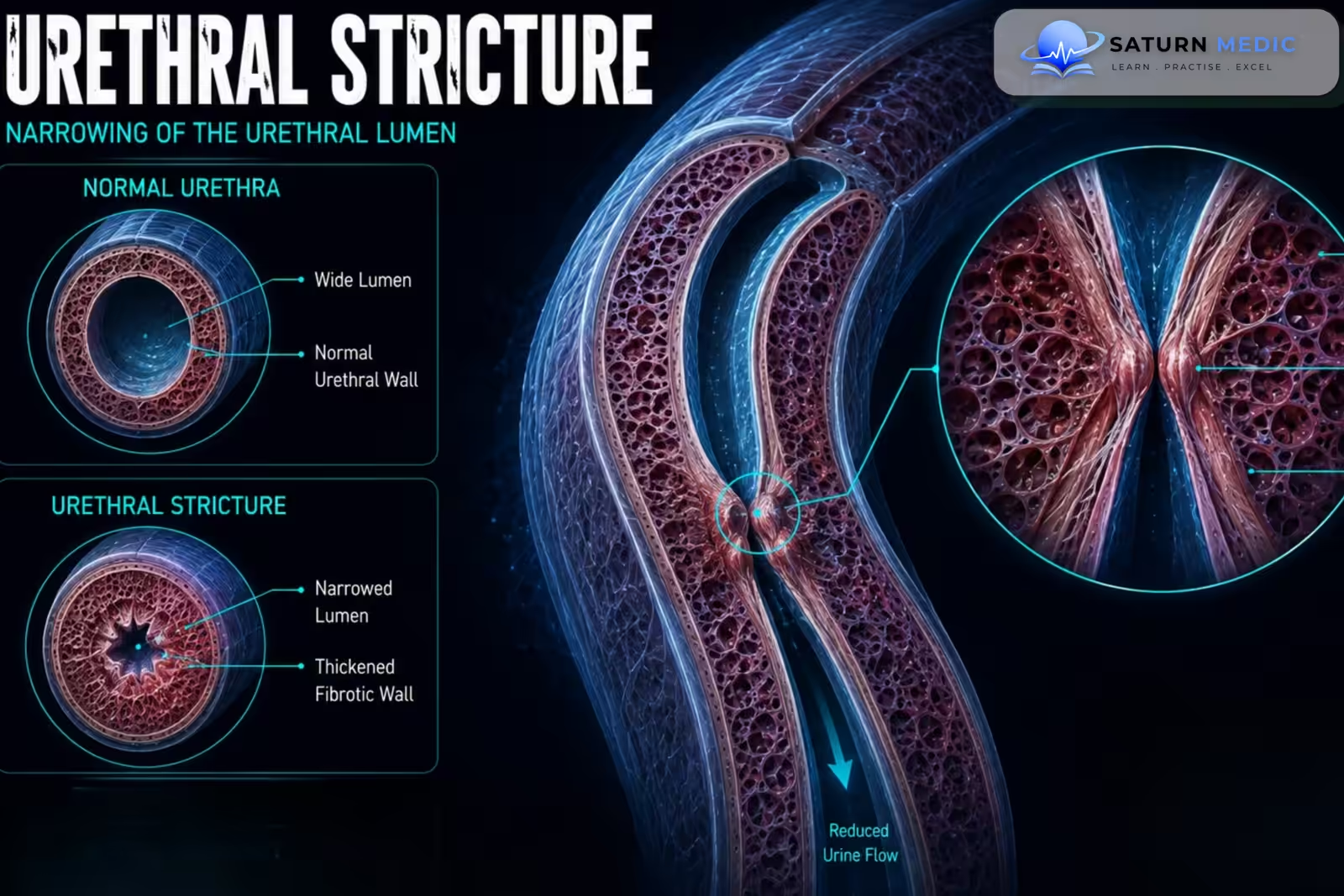
Urethral Stricture is an abnormal narrowing of the urethra caused by scarring and fibrosis of the urethral tissue. The narrowing restricts the normal flow of urine and can lead to weak urinary stream, urinary retention, recurrent infection, bladder dysfunction, and kidney damage if severe or untreated
Also called
ICD-10
Specialty
Onset
Reviewed
-
OverviewOverview
-
Etiology & Risk FactorsEtiology & Risk Factors
-
PathophysiologyPathophysiology
-
Clinical PresentationClinical Presentation
-
History TakingHistory Taking
-
Physical ExaminationPhysical Examination
-
InvestigationsInvestigations
-
DiagnosisDiagnosis
-
ManagementManagement
-
ComplicationsComplications
-
PrognosisPrognosis
-
Key Points / Clinical PearlsKey Points / Clinical Pearls
Overview
–Urethral Stricture occurs when inflammation, trauma, or injury causes scar tissue to form within or around the urethra. The scar contracts over time, reducing the diameter of the urethral lumen.
The condition may affect:
- Anterior urethra: including the penile and bulbar urethra
- Posterior urethra: usually associated with pelvic trauma or previous treatment
- A short or long segment of the urethra
-The severity depends on the location, length, degree of narrowing, and underlying cause.
Etiology & Risk Factors
-Etiology
The main causes of Urethral Stricture are:
- Previous urethral trauma
- Pelvic fracture-associated urethral injury
- Straddle injury
- Urethral catheterization or instrumentation
- Previous urethral surgery
- Infection or chronic inflammation
- Lichen sclerosus
- Previous radiation therapy
- Idiopathic causes
-Risk Factors
- Previous Urethral Trauma
- Repeated urethral catheterization
- Previous endoscopic procedures
Pathophysiology
Urethral injury or inflammation → tissue damage → healing with collagen deposition → fibrosis and scar formation → progressive narrowing of the urethral lumen → increased resistance to urine flow → bladder outlet obstruction → urinary retention, infection, and upper urinary tract damage
Clinical Presentation
–Symptoms
-
Symptoms usually develop gradually but may become acute if obstruction becomes severe.
- Weak urinary stream
- Straining to void
- Hesitancy
- Intermittent urine flow
- Prolonged urination
- Incomplete bladder emptying
- Post-void dribbling
- Urinary frequency
-Signs:
- Reduced urinary stream
- Palpable distended bladder in retention
- Suprapubic discomfort
- Perineal scarring in some patients
- Evidence of previous trauma or surgery
History Taking
-Ask about:
- Weak or spraying urinary stream
- Difficulty starting urination
- Straining or prolonged voiding
- Incomplete emptying
- Post-void dribbling
- Episodes of urinary retention
- Recurrent Urinary Tract Infection (UTI)
- Hematuria
- Previous pelvic, perineal, or penile trauma
- Previous urethral catheterization
Physical Examination
-General Examination
- Assess vital signs if infection or urinary retention is present
- Assess for suprapubic pain or discomfort
- Look for signs of renal impairment in advanced obstruction
-System-Specific Examination:
- Palpate for a distended bladder
- Inspect the external genitalia and urethral meatus
- Look for penile or perineal scars
- Examine for signs of lichen sclerosus when clinically suspected
Investigations
Biochemistry / Specific Tests
-
Urinalysis
-
Urine culture when infection is suspected
-
Serum creatinine and electrolytes in significant or prolonged obstruction
-
Uroflowmetry to assess urinary flow rate
-
Post-void residual urine measurement
A reduced maximum urinary flow with a prolonged, flattened flow pattern supports bladder outlet obstruction but does not confirm the cause.
-Imaging
Retrograde Urethrography
Retrograde urethrography is one of the key investigations for defining Urethral Stricture.
It assesses:
-
Location
-
Length
-
Number of strictures
-
Severity of narrowing
-Voiding Cystourethrography
May be combined with retrograde imaging, particularly for complex or posterior strictures.
Special / Confirmatory Tests
-Cystoscopy
Cystoscopy allows direct visualization of the stricture and may help assess the urethra and bladder.
-Urethral Ultrasound
May be used in selected cases to assess stricture length and surrounding scar tissue.
Diagnosis
-The diagnosis of Urethral Stricture is based on:
Typical obstructive urinary symptoms → reduced urinary flow on uroflowmetry → imaging or endoscopic demonstration of urethral narrowing.
-Retrograde urethrography is especially important for defining the anatomical characteristics before reconstructive treatment.
Management
1. First-Line / Emergency Management
Acute urinary retention requires prompt bladder drainage.
Management may include:
-
Careful urethral catheterization by an experienced clinician when appropriate
-
Suprapubic catheterization if urethral catheterization is unsuccessful or unsafe
-
Treatment of associated infection
-
Analgesia when required
2. Definitive Treatment
The definitive treatment depends on the stricture’s length, location, cause, and previous treatment.
-Dilation
Gradual mechanical dilation may be used for selected short strictures.
-Direct Vision Internal Urethrotomy
An endoscopic incision is made through the stricture to widen the urethral lumen. It is mainly suitable for selected short strictures but recurrence is common.
-Urethroplasty
Urethroplasty is the definitive reconstructive treatment for many recurrent, long, or complex strictures.
-Techniques include:
-
Excision and primary anastomosis for selected short strictures
-
Substitution urethroplasty using tissue grafts, commonly buccal mucosa
3. Medical Treatment
There is no medication that permanently removes established urethral scar tissue.
Medical treatment may include:
-
Antibiotics for confirmed infection
-
Treatment of underlying inflammatory disease
-
Management of associated bladder symptoms when appropriate
Complications
- Acute urinary retention
- Recurrent Urinary Tract Infection (UTI)
- Bladder stones
- Bladder diverticula
- Periurethral abscess
- Urethral fistula
- Hydronephrosis
- Acute Kidney Injury (AKI)
Prognosis
The prognosis depends mainly on the length, location, cause, severity, and treatment history of the stricture. Short, uncomplicated strictures may respond to endoscopic treatment, although recurrence is common. Long, recurrent, or complex strictures generally have better long-term outcomes after appropriate urethroplasty. Untreated severe obstruction can cause progressive bladder dysfunction and kidney damage.
Key Points / Clinical Pearls
- Urethral Stricture is narrowing of the urethra caused by fibrosis and scar formation.
- Previous trauma and urethral instrumentation are important causes.
- A weak urinary stream is a common presenting symptom.
- Hesitancy, straining, and post-void dribbling may occur.
- Recurrent Urinary Tract Infection (UTI) can result from poor bladder emptying.
- Acute urinary retention may require urgent bladder drainage.
- Uroflowmetry helps identify obstructed urinary flow.
- Post-void residual urine helps assess incomplete bladder emptying.
- Retrograde urethrography defines the location and length of the stricture.
- Cystoscopy can directly visualize the narrowed segment.
- European Association of Urology (EAU). EAU Guidelines on Urethral Strictures .
- American Urological Association (AUA). Urethral Stricture Guideline .
- Wessells H, Angermeier KW, Elliott S, et al. Male Urethral Stricture: American Urological Association Guideline. J Urol. 2017;197(1):182-190. Journal of Urology .
- Mundy AR, Andrich DE. Urethral Strictures. BJU Int. 2011;107(1):6-26. BJU International .
- Santucci RA, Joyce GF, Wise M. Male Urethral Stricture Disease. J Urol. 2007;177(5):1667-1674. PubMed .
- National Library of Medicine (NIH). Urethral Strictures . StatPearls.