

Clinical Subject Page
Bladder Cancer
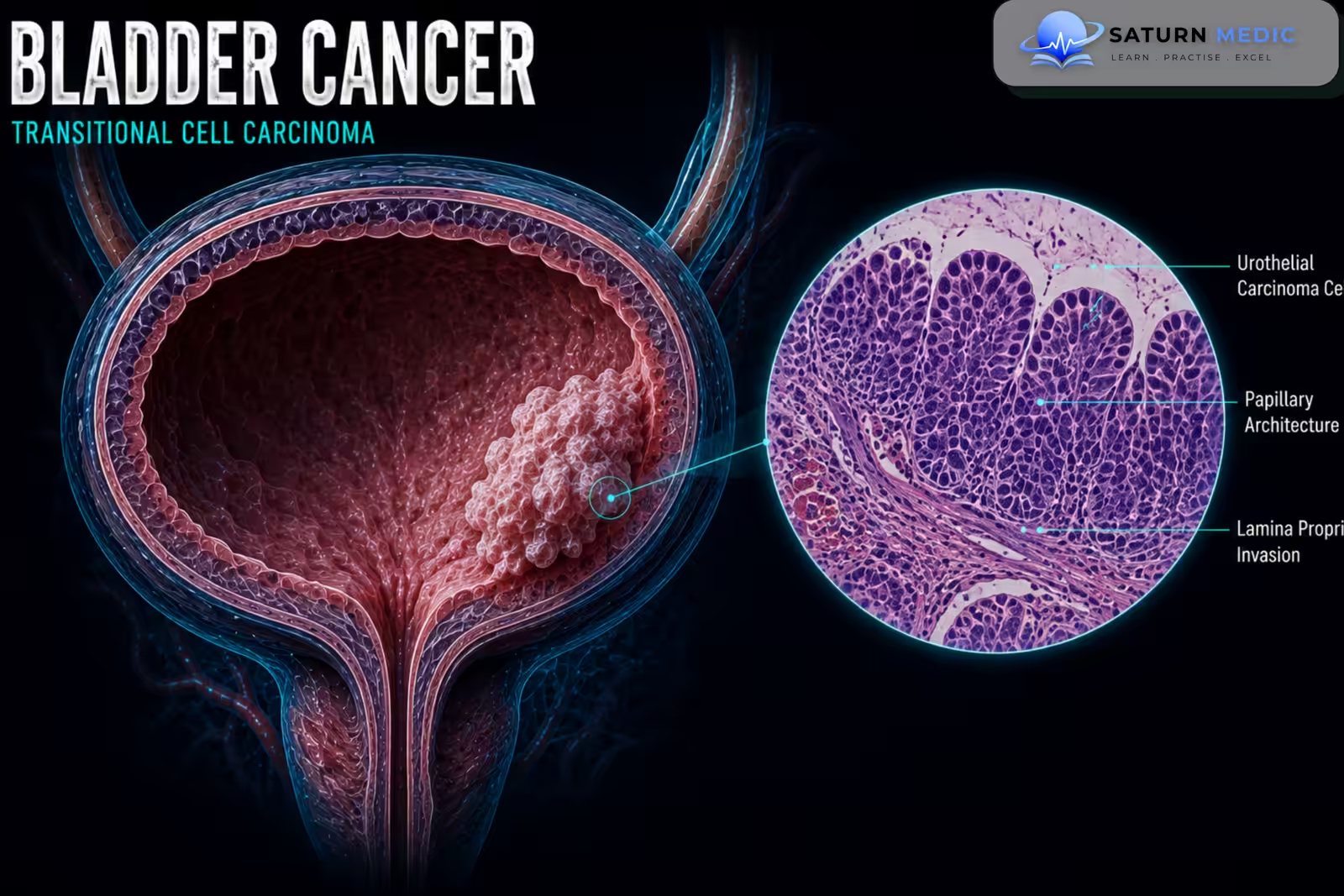
Bladder Cancer is a malignant tumor arising from the tissues of the urinary bladder. Most cases are urothelial carcinoma, which develops from the urothelial lining of the bladder. Bladder Cancer ranges from superficial non-muscle-invasive disease to aggressive muscle-invasive and metastatic disease
Also called
ICD-10
Specialty
Onset
Reviewed
-
OverviewOverview
-
Etiology & Risk FactorsEtiology & Risk Factors
-
PathophysiologyPathophysiology
-
Clinical PresentationClinical Presentation
-
History TakingHistory Taking
-
Physical ExaminationPhysical Examination
-
InvestigationsInvestigations
-
DiagnosisDiagnosis
-
ManagementManagement
-
ComplicationsComplications
-
PrognosisPrognosis
-
Key Points / Clinical PearlsKey Points / Clinical Pearls
Overview
-MostBladder Cancer is urothelial carcinoma. Clinically, it is broadly classified into:
- Non-muscle-invasive Bladder Cancer (NMIBC): confined to the mucosa or lamina propria.
- Muscle-invasive Bladder Cancer (MIBC): invades the detrusor muscle or beyond.
- Metastatic Bladder Cancer: spreads to distant organs.
This classification is important because treatment and prognosis differ substantially between stages.
Etiology & Risk Factors
-Etiology
–Bladder Cancer develops through genetic and molecular changes in urothelial cells, often following prolonged exposure to urinary carcinogens.
Most tumors are urothelial carcinomas.
-Risk Factors
- Cigarette smoking
- Occupational exposure to aromatic amines and certain industrial chemicals
- Increasing age
- Chronic bladder irritation or inflammation
- Previous pelvic radiotherapy
Pathophysiology
Exposure to urinary carcinogens → urothelial DNA damage → accumulation of genetic mutations → uncontrolled urothelial cell proliferation → tumor formation → local invasion → lymphatic and hematogenous spread in advanced disease
Clinical Presentation
-Symptoms:
- Painless visible hematuria
- Microscopic hematuria
- Dysuria
- Urinary frequency
- Urinary urgency
- Pelvic pain in advanced disease
- Flank pain due to ureteric obstruction
-Signs:
Early Bladder Cancer may have no specific physical signs.
Possible findings in advanced disease include:
- Palpable pelvic mass
- Weight loss
- Lower-limb edema
- Supraclavicular or other lymphadenopathy
History Taking
-Ask about:
- Visible or microscopic hematuria
- Whether hematuria is painful or painless
- Dysuria, frequency, and urgency
- Flank or pelvic pain
- Smoking history
- Occupational chemical exposure
- Previous pelvic radiotherapy
- Recurrent Urinary Tract Infection (UTI)
- Previous bladder tumors
Physical Examination
-General Examination
- Assess general condition and weight loss
- Check for peripheral edema
- Assess for lymphadenopathy when advanced disease is suspected
-System-Specific Examination:
- Examine the abdomen for masses
- Assess for suprapubic tenderness or a palpable bladder
- Perform a focused pelvic examination when clinically indicated
Investigations
Biochemistry / Specific Tests
Urinalysis
Urine culture when infection is suspected
Renal function assessment
Urine cytology in selected patients, particularly when high-grade disease is suspected
-Imaging
Computed Tomography Urography (CT Urography)
Used to assess the urinary tract and identify:
Bladder masses
Upper urinary tract lesions
Hydronephrosis
Regional or distant disease
-Special / Confirmatory Tests
Cystoscopy
Direct visualization of the bladder and detection of suspicious lesions.
Transurethral Resection of Bladder Tumor (TURBT)
Provides tissue for histological diagnosis and determines tumor stage and grade.
-Important Investigation Note
Cystoscopy with TURBT is central to the diagnosis and initial management of suspected Bladder Cancer.
Diagnosis
-Bladder Cancer is diagnosed by:
Hematuria or suspicious urinary symptoms → cystoscopy → identification of bladder lesion → TURBT and histological examination → tumor staging and grading.
Imaging is used to assess the upper urinary tract and stage more advanced disease.
Management
Bladder Cancer · Management Overview
Bladder Cancer · Surgical Management
- Transurethral resection of bladder tumour
- 1st-line for all stages — diagnostic + therapeutic
- Re-TURBT at 4–6 weeks if T1 or incomplete resection
- Photodynamic / NBI guidance improves detection
- Standard of care for MIBC (T2–T4a)
- Removes bladder + prostate/uterus/anterior vaginal wall
- Bilateral pelvic lymph node dissection mandatory
- Open, laparoscopic, or robotic (RARC) approaches
- Bladder-sparing alternative to cystectomy
- Maximal TURBT → concurrent chemoradiation
- Cisplatin or 5-FU/MMC as radiosensitiser
- Salvage cystectomy if residual/recurrent MIBC
- Rare — only solitary tumour at dome/wall, away from trigone
- Adequate surgical margins required
- High local recurrence risk — use selectively
- Low risk: cystoscopy at 3 months → then annually × 5 yrs
- High risk: every 3 months × 2 yrs → every 6 months × 3 yrs
- Urine cytology at each visit
- Performed with radical cystectomy
- Extended template preferred — ↑ staging accuracy + OS
- Removes: obturator, external/internal iliac, presacral nodes
Bladder Cancer · Urinary Diversion After Cystectomy
- Ureters → isolated ileal segment → stoma (right lower abdomen)
- Urine drains continuously into external bag
- Simplest, shortest operative time — preferred in elderly/comorbid
- No need for catheterisation
- Ileum fashioned into reservoir → anastomosed to urethra
- Patient voids normally — no stoma, no bag
- Requires intact urethra + sphincter; not for urethral involvement
- Risk: nocturnal incontinence, urinary retention (self-catheterise)
- Pouch constructed from bowel → continent stoma on abdomen
- Patient self-catheterises 4–6× daily — no external bag
- Indiana pouch / Kock pouch most common types
- Used when urethra cannot be preserved
Complications
- Recurrent tumor
- Progression to muscle-invasive disease
- Metastatic disease
- Ureteric obstruction
- Hydronephrosis
- Kidney dysfunction
- Severe hematuria
- Anemia
- Urinary obstruction
Prognosis
The prognosis of Bladder Cancer depends mainly on tumor stage, grade, presence of carcinoma in situ, recurrence, progression, and metastatic spread. Non-muscle-invasive disease generally has a better prognosis but often recurs, while muscle-invasive and metastatic disease carry a significantly worse prognosis
Key Points / Clinical Pearls
- Bladder Cancer is most commonly urothelial carcinoma.
- Painless visible hematuria is the classic presentation.
- Smoking is the most important modifiable risk factor.
- Occupational chemical exposure can increase risk.
- Hematuria may be intermittent.
- Cystoscopy is central to diagnosis.
- TURBT provides histological diagnosis and initial staging.
- NMIBC is confined to the mucosa or lamina propria.
- MIBC invades the detrusor muscle.
- Intravesical BCG is used for appropriate high-risk NMIBC.
- Radical cystectomy is a major treatment for MIBC.
- Bladder-preserving multimodal treatment is possible in selected patients.
- Bladder Cancer has a high recurrence rate.
- European Association of Urology (EAU). EAU Guidelines on Non-Muscle-Invasive Bladder Cancer.
- European Association of Urology (EAU). EAU Guidelines on Muscle-Invasive and Metastatic Bladder Cancer.
- American Urological Association (AUA). Non-Muscle Invasive Bladder Cancer Guideline.
- National Comprehensive Cancer Network (NCCN). NCCN Guidelines for Bladder Cancer.
- National Cancer Institute (NCI). Bladder Cancer.