

Clinical Subject Page
Barrett Esophagus
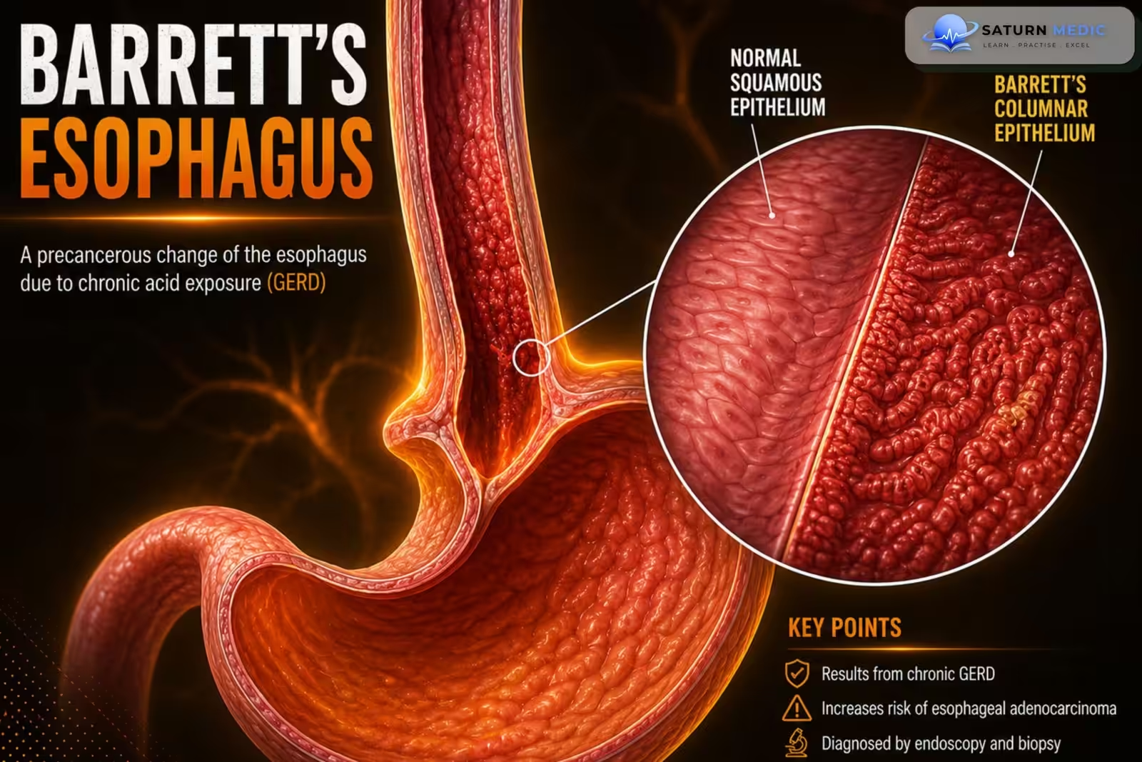
Barrett esophagus is a precancerous condition in which the normal stratified squamous epithelium
of the distal esophagus is replaced by nonciliated columnar epithelium containing goblet cells
(intestinal metaplasia).
Also called
ICD-10
Specialty
Onset
Reviewed
-
OverviewOverview
-
Etiology & Risk FactorsEtiology & Risk Factors
-
PathophysiologyPathophysiology
-
Clinical PresentationClinical Presentation
-
History TakingHistory Taking
-
Physical ExaminationPhysical Examination
-
InvestigationsInvestigations
-
DiagnosisDiagnosis
-
ManagementManagement
-
ComplicationsComplications
-
PrognosisPrognosis
-
Key Points / Clinical PearlsKey Points / Clinical Pearls
Overview
Barrett esophagus develops mainly as a result of chronic gastroesophageal reflux disease (GERD).
It is a precursor lesion for esophageal adenocarcinoma. Diagnosis requires endoscopy with biopsy
demonstrating intestinal metaplasia. Management includes daily proton pump inhibitor (PPI)
therapy, surveillance endoscopy, and endoscopic eradication therapy for dysplasia.
Etiology & Risk Factors
• Chronic gastroesophageal reflux disease (GERD) (most common cause)
• Male sex
• Age >50 years
• Obesity
• Smoking
• Family history of Barrett esophagus
Pathophysiology
1. Chronic acid reflux injures the distal esophageal mucosa.
2. Normal stratified squamous epithelium is replaced by nonciliated columnar epithelium with goblet
cells (intestinal metaplasia).
3. Barrett esophagus becomes a precursor lesion for esophageal adenocarcinoma.
4. Dysplasia may develop before progression to invasive cancer
Clinical Presentation
Barrett esophagus itself usually does not cause symptoms.
Patients may have symptoms of underlying GERD, including:
• Heartburn
• Regurgitation
History Taking
Ask about:
• History of chronic GERD
• Heartburn
• Regurgitation
• Duration of reflux symptoms
• Risk factors: Age >50 years, Male sex, Obesity, Smoking, Family history of Barrett esophagus
Physical Examination
Physical examination is often normal.
Evaluate for signs related to GERD or possible complications if present.
Investigations
Esophagogastroduodenoscopy (EGD)
– Salmon-pink mucosa extending proximal to the gastroesophageal junction (GEJ)Esophageal biopsy
– Confirms intestinal metaplasia on histopathology
Diagnosis
Diagnosis is usually based on:
• Characteristic endoscopic findings on EGD
• Histopathological confirmation of intestinal metaplasia from biopsy
Related Topics
Management
1. Control Gastroesophageal Reflux
Give a proton pump inhibitor (PPI), usually once daily.
Increase to twice daily when reflux symptoms remain uncontrolled or during endoscopic treatment when indicated.
Encourage:
Weight reduction when overweight
Smoking cessation
Avoiding late meals
Elevating the head of the bed for nocturnal reflux
Avoiding individual reflux triggers
Antireflux surgery is considered for persistent reflux despite optimized medical therapy, but it is not recommended solely to prevent cancer.
2. Endoscopic Surveillance
Non-dysplastic Barrett’s Esophagus
No immediate ablation is required.
Continue PPI therapy.
Perform periodic surveillance upper endoscopy with systematic biopsies.
Surveillance intervals are usually based on the length of the Barrett’s segment:
<3 cm: approximately every 5 years
≥3 cm: approximately every 3 years
Indefinite for Dysplasia
Confirm the diagnosis with an expert gastrointestinal pathologist.
Optimize acid suppression, often with twice-daily PPI.
Repeat endoscopy with biopsies after approximately 6 months.
Persistent indefinite dysplasia requires closer surveillance.
3. Low-Grade Dysplasia
Confirm the diagnosis by a second expert gastrointestinal pathologist.
Endoscopic eradication therapy is generally preferred.
Endoscopic surveillance remains an alternative in selected patients after discussing risks and benefits.
Common eradication treatment:
Endoscopic resection of visible lesions
Radiofrequency ablation of the remaining Barrett’s mucosa
4. High-Grade Dysplasia
Refer to an experienced specialist or high-volume center.
Perform careful endoscopic assessment.
Remove visible or nodular lesions using:
Endoscopic mucosal resection
Endoscopic submucosal dissection in selected cases
Ablate the remaining Barrett’s epithelium, commonly with radiofrequency ablation.
High-grade dysplasia should generally receive endoscopic eradication therapy because of its substantial risk of progression to adenocarcinoma.
5. Early Esophageal Adenocarcinoma
For superficial cancer limited to the mucosa:
Endoscopic resection of the cancer
Ablation of the remaining Barrett’s mucosa
Close surveillance afterward
Esophagectomy may be required when there is:
Deep submucosal invasion
Lymphovascular invasion
Poor differentiation
Positive deep resection margins
Suspected lymph-node involvement
Disease unsuitable for endoscopic treatment
6. Surveillance After Eradication
Even after complete eradication of intestinal metaplasia:
Continue PPI therapy.
Perform scheduled surveillance endoscopy.
Carefully inspect the gastroesophageal junction and previous Barrett’s segment.
Biopsy suspicious areas because recurrence can occur.
Complications
• Dysplasia
• Esophageal adenocarcinoma
Prognosis
- Most patients remain stable with appropriate acid suppression and surveillance.
- The major concern
is progression to dysplasia and esophageal adenocarcinoma, making regular follow-up essential
Key Points / Clinical Pearls
• Barrett esophagus is caused mainly by chronic GERD.
• It is characterized by intestinal metaplasia of the distal esophagus.
• It is a precursor lesion for esophageal adenocarcinoma.
• Barrett esophagus itself is usually asymptomatic; symptoms are typically due to GERD.
• Diagnosis requires EGD with biopsy.
• Daily PPI therapy is recommended for all patients.
• Surveillance endoscopy is required for patients without dysplasia.
• Dysplasia should be treated with endoscopic eradication therapy
- National Center for Biotechnology Information (NIH). Barrett Esophagus, StatPearls.
- Wani S, Zhou MJ, Sawas T, et al. AGA Clinical Practice Guideline on Surveillance of Barrett's Esophagus. Gastroenterology. 2025;169:1184-1231. PMID: 41125322.
- Shaheen NJ, Falk GW, Iyer PG, et al. Diagnosis and Management of Barrett's Esophagus: An Updated ACG Guideline. Am J Gastroenterol. 2022;117:559-587. PMID: 35354777.
- MedlinePlus, National Library of Medicine (NIH). Barrett Esophagus: Medical Encyclopedia.
- National Center for Biotechnology Information (NIH). Esophageal Adenocarcinoma, StatPearls.