

Clinical Subject Page
Boerhaave Syndrome
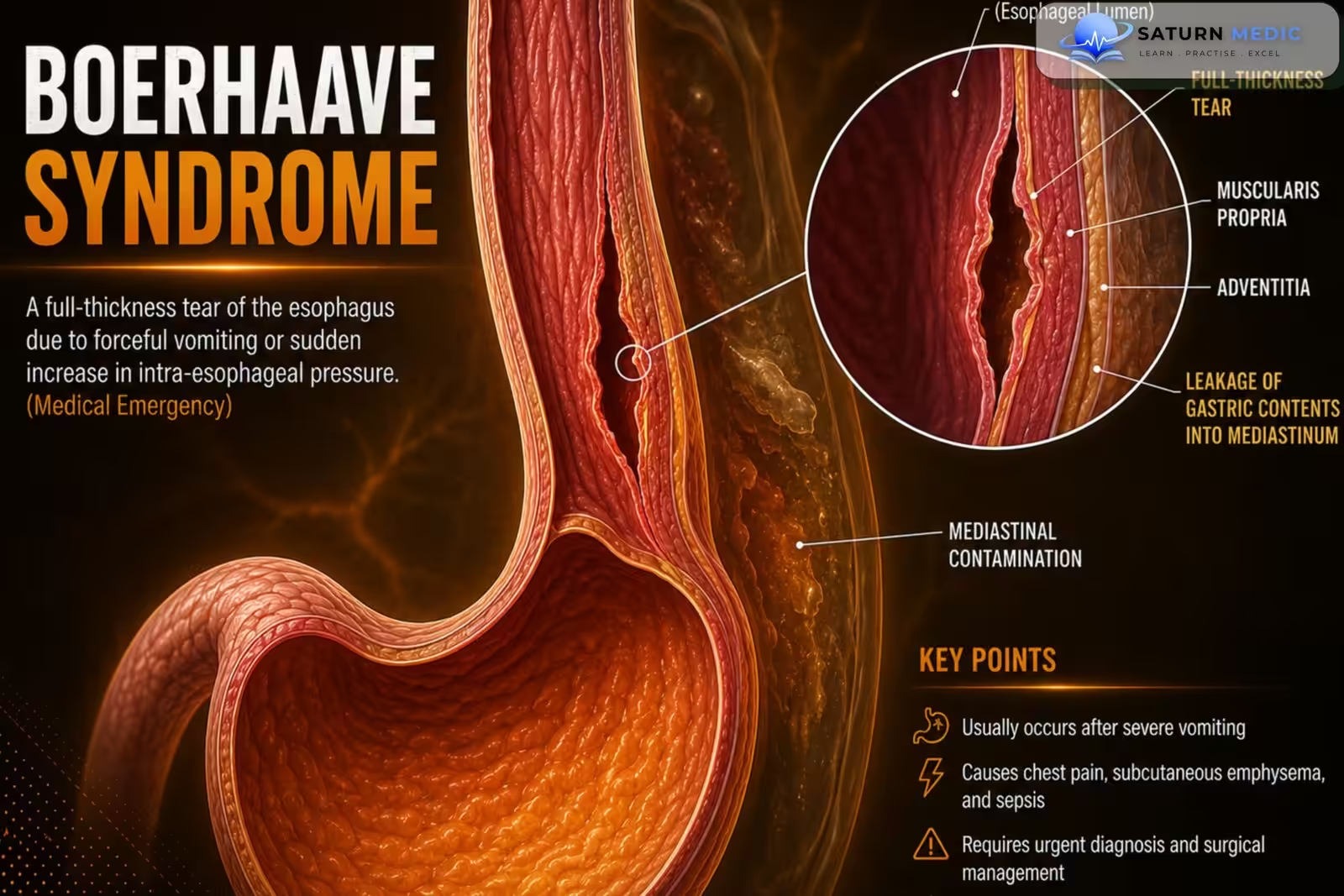
Boerhaave syndrome is a spontaneous transmural (full-thickness) rupture of the esophagus,
usually following forceful vomiting or retching, resulting in leakage of esophageal contents into the
mediastinum
Also called
ICD-10
Specialty
Onset
Reviewed
-
OverviewOverview
-
Etiology & Risk FactorsEtiology & Risk Factors
-
PathophysiologyPathophysiology
-
Clinical PresentationClinical Presentation
-
History TakingHistory Taking
-
Physical ExaminationPhysical Examination
-
InvestigationsInvestigations
-
DiagnosisDiagnosis
-
ManagementManagement
-
ComplicationsComplications
-
PrognosisPrognosis
-
Key Points / Clinical PearlsKey Points / Clinical Pearls
Overview
Boerhaave syndrome is a life-threatening subtype of esophageal perforation caused by a sudden increase in intrathoracic pressure during forceful vomiting. It most commonly affects the distal third
of the esophagus on the left dorsolateral wall. Prompt diagnosis and treatment are essential to prevent mediastinitis, sepsis, and death.
Etiology & Risk Factors
• Repeated forceful vomiting or retching
• Recent large intake of alcohol
• Recent large meal
• Prolonged coughing
• Childbirth
• Seizures
• Weightlifting
Pathophysiology
1. Forceful vomiting causes a marked increase in intrathoracic pressure.
2. A full-thickness (transmural) tear develops in the esophageal wall.
3. In over 90% of cases, the rupture occurs in the distal third of the esophagus on the left
dorsolateral wall.
4. Esophageal contents leak into the mediastinum.
5. This can rapidly lead to mediastinitis, sepsis, and multiorgan dysfunction
Clinical Presentation
Classic Mackler triad:
• Vomiting or retching
• Severe retrosternal chest pain (often radiating to the back)
• Subcutaneous or mediastinal emphysema
Other features:
• Dyspnea
• Tachypnea
• Tachycardia
• Dysphagia
• Signs of sepsis
• Delayed presentation may include multiorgan dysfunction
History Taking
-
Ask about:
• Recent forceful vomiting or retching
• Heavy alcohol intake
• Large meal before symptom onset
• Sudden severe chest pain
• Pain radiating to the back
• Dysphagia
• Dyspnea
• Recent upper endoscopy
• Trauma
• Symptoms of sepsis (fever, chills)
Physical Examination
Assess for:
• Subcutaneous emphysema (neck or suprasternal crepitus)
• Hamman sign (mediastinal crunch synchronous with the heartbeat)
• Tachycardia
• Tachypnea
• Signs of sepsis
• Hemodynamic instability
Investigations
-Initial imaging:
• Chest X-ray
• Upright abdominal X-ray (if indicated)
• Neck X-ray (if cervical perforation is suspected)
-Possible findings:
• Pneumomediastinum
• Pneumothorax
• Pleural effusion
• Subcutaneous emphysema
• Widened mediastinum
• Pneumoperitoneum
• Mediastinal air-fluid levels
-Confirmatory tests:
• Contrast esophagography (gold standard)
• CT chest with CT esophagography (oral contrast when feasible)
-Endoscopy:
• Flexible endoscopy may be used in selected situations but is generally avoided in nonpenetrating
injuries unless there is a specific therapeutic indication.
Diagnosis
-
Diagnosis is usually based on:
• Clinical suspicion
• Initial radiographic imaging
• Confirmation with contrast esophagography and/or CT esophagography
Related Topics
Management
-
1. Initial Stabilization
-
Assess Airway, Breathing, and Circulation (ABC)
-
Oxygen therapy
-
Establish large-bore IV access
-
Intravenous fluid resuscitation
-
Hemodynamic monitoring
-
Keep the patient nil by mouth (NPO)
2. Medical Therapy
-
Broad-spectrum intravenous antibiotics
-
Intravenous proton pump inhibitor (PPI)
-
Adequate analgesia
-
Antiemetics
-
Nutritional support (enteral or parenteral if prolonged NPO)
3. Definitive Treatment
Surgical Repair (Preferred)
Indicated for:
-
Early presentation (<24 hours)
-
Large perforation
-
Sepsis
-
Mediastinitis
-
Hemodynamic instability
Options:
-
Primary esophageal repair
-
Debridement of contaminated tissue
-
Mediastinal and pleural drainage
Endoscopic Management
Selected stable patients with:
-
Small, contained perforations
-
Minimal contamination
Options:
-
Endoscopic stent placement
-
Endoscopic clips or endoscopic vacuum therapy (selected cases)
Conservative Management
Consider only in carefully selected patients with:
-
Small contained leak
-
No sepsis
-
Hemodynamic stability
-
Minimal mediastinal contamination
Management includes:
-
NPO
-
IV antibiotics
-
IV fluids
-
Close monitoring
-
Nutritional support
4. Follow-Up
-
Repeat contrast esophagram or CT with oral contrast to confirm healing before resuming oral intake
-
Monitor for infection, mediastinitis, and leak recurrence
-
Gradual return to oral feeding after healing is confirmed
-
Complications
• Mediastinitis
• Sepsis
• Multiorgan dysfunction
• Pneumomediastinum
• Pneumothorax
• Pleural effusion
Prognosis
- Prognosis depends on early recognition and prompt treatment.
- Delayed diagnosis increases the
risk of mediastinitis, sepsis, multiorgan failure, and mortality.
Key Points / Clinical Pearls
• Boerhaave syndrome is a spontaneous full-thickness esophageal rupture.
• It is most commonly caused by forceful vomiting or retching.
• Heavy alcohol intake and large meals are common risk factors.
• The classic Mackler triad consists of vomiting, severe chest pain, and subcutaneous emphysema.
• Chest X-ray is the initial imaging study.
• Contrast esophagography is the gold-standard confirmatory test.
• CT chest with CT esophagography is useful when the diagnosis is uncertain or the patient is
unstable.
• Surgical repair is the usual treatment.
• Delayed diagnosis greatly increases morbidity and mortality.
- Turner AR, Collier SA, Turner SD. National Center for Biotechnology Information (NIH). Boerhaave Syndrome, StatPearls.
- National Center for Biotechnology Information (NIH). Esophageal Perforation and Tears, StatPearls.
- Kuwano H, Yokobori T, Kumakura Y, et al. Pathophysiology of Vomiting and Esophageal Perforation in Boerhaave's Syndrome. Dig Dis Sci. 2020;65:3253-3259. PMID: 31965389.
- MedlinePlus, National Library of Medicine (NIH). Esophageal Rupture: Medical Encyclopedia.
- Biancari F, D'Andrea V, Paone R, et al. Current Treatment and Outcome of Esophageal Perforations in Adults: Systematic Review and Meta-Analysis of 75 Studies. World J Surg. 2013;37:1051-1059. PMID: 23404161.