

Clinical Subject Page
Chronic Gastritis
Zollinger-Ellison syndrome is a gastrin-secreting neuroendocrine tumor (gastrinoma), most
commonly located in the duodenum or pancreas, causing excessive gastric acid secretion.
Also called
ICD-10
Specialty
Onset
Reviewed
-
OverviewOverview
-
Etiology & Risk FactorsEtiology & Risk Factors
-
PathophysiologyPathophysiology
-
Clinical PresentationClinical Presentation
-
History TakingHistory Taking
-
Physical ExaminationPhysical Examination
-
InvestigationsInvestigations
-
DiagnosisDiagnosis
-
ManagementManagement
-
ComplicationsComplications
-
PrognosisPrognosis
-
Key Points / Clinical PearlsKey Points / Clinical Pearls
Overview
H. pylori is a gram-negative bacterium that colonizes the stomach. Infection usually occurs in
childhood and persists for life if untreated. Although many patients are asymptomatic, H. pylori
infection is strongly associated with chronic gastritis, peptic ulcer disease, gastric cancer, and
gastric MALT lymphoma. Diagnosis is made using noninvasive testing or gastric biopsy, and all
infected patients should receive eradication therapy.
Etiology & Risk Factors
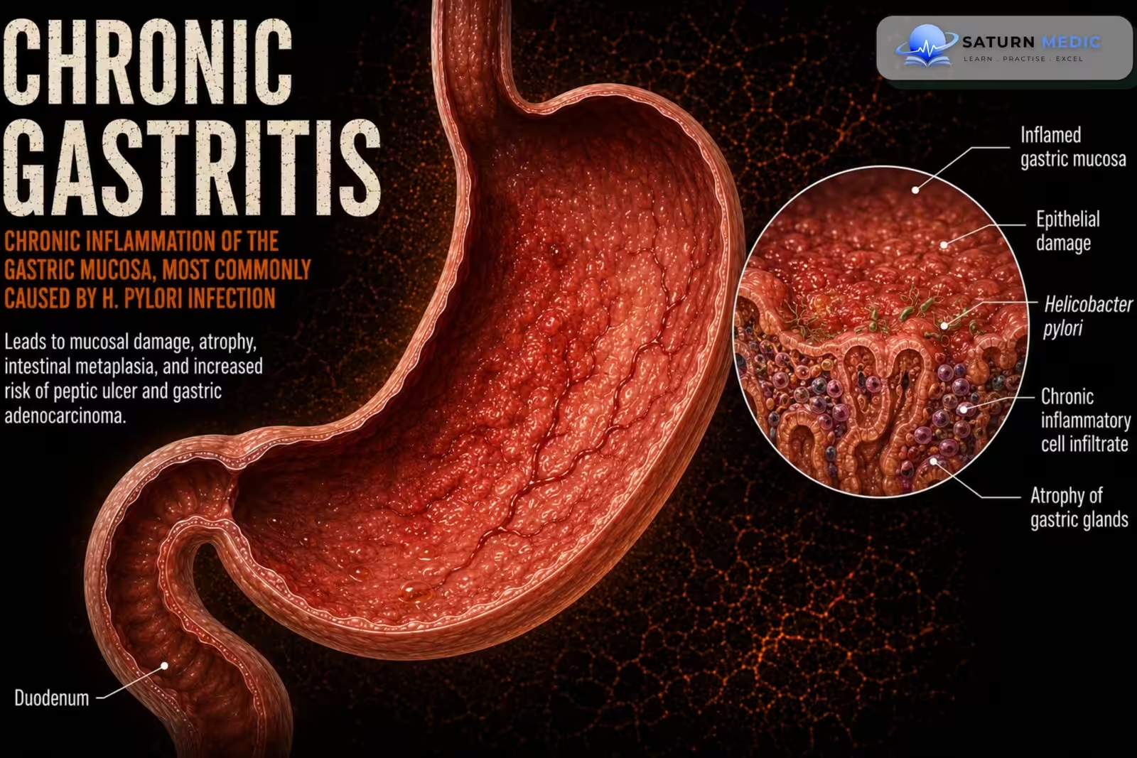
Chronic gastritis is a persistent inflammation of the gastric mucosa. Helicobacter pylori (H. pylori) infection is the most common cause worldwide. The bacterium colonizes the gastric mucosa, leading to chronic inflammation, mucosal injury, and, over time, gastric atrophy and intestinal metaplasia. Less commonly, chronic gastritis may result from autoimmune disease, prolonged NSAID use, bile reflux, or other infections.
Risk Factors
- Helicobacter pylori infection
- Living in crowded or low-sanitation environments
- Family history of H. pylori infection or gastric cancer
- Increasing age
- Smoking
- Long-term NSAID use
- Excessive alcohol consumption
- Autoimmune disorders
- Low socioeconomic status
Pathophysiology
1. H. pylori colonizes the gastric mucosa.
2. Chronic inflammation develops with a predominantly mononuclear inflammatory infiltrate.
3. Progressive loss of normal gastric gland architecture occurs.
4. Persistent infection increases the risk of peptic ulcer disease, gastric cancer, and MALT
lymphoma.
Clinical Presentation
Common Symptoms
-
Epigastric pain or burning
-
Dyspepsia (indigestion)
-
Early satiety
-
Postprandial fullness
-
Nausea
-
Bloating
-
Belching
-
Mild upper abdominal discomfort
-
Loss of appetite
-
Many patients are asymptomatic
Five Important Symptoms That Are Often Missed
-
Iron-deficiency anemia (fatigue, weakness)
-
Vitamin B12 deficiency (paresthesia, numbness, glossitis) due to chronic atrophic gastritis
-
Unintentional weight loss
-
Occult gastrointestinal bleeding (positive fecal occult blood test or chronic anemia)
-
Halitosis (bad breath) associated with chronic H. pylori infection
History Taking
-
Ask about:
• Dyspepsia
• Epigastric pain
• Duration of symptoms
• Previous diagnosis or treatment for H. pylori
• Previous peptic ulcer disease
• Family or personal history of gastric cancer (if relevant)
Physical Examination
Physical examination is often normal.
Assess for epigastric tenderness and features of complications if suspected.
Investigations
-
Initial Laboratory Tests
-
Complete blood count (CBC)
-
Iron studies (if anemia is suspected)
-
Vitamin B12 level (if atrophic gastritis is suspected)
-
Stool occult blood test (if gastrointestinal bleeding is suspected)
Diagnostic Investigations
H. pylori Testing
-
Urea breath test (preferred non-invasive test)
-
Stool H. pylori antigen test
-
Rapid urease test (during endoscopy)
-
Histology from gastric biopsy
-
Culture or PCR (selected cases)
Upper Gastrointestinal Endoscopy (EGD)
Performed in patients with:
-
Alarm symptoms
-
Older age at presentation
-
Suspected gastric malignancy
-
Failure of initial treatment
May demonstrate:
-
Gastric erythema
-
Mucosal atrophy
-
Nodularity
-
Erosions or ulcers
Biopsy confirms chronic gastritis and detects H. pylori, intestinal metaplasia, dysplasia, or malignancy.
Additional Investigations
-
Histopathological examination of gastric biopsies
-
Serum gastrin and autoimmune antibody testing (if autoimmune gastritis is suspected)
-
Diagnosis
Diagnosis is confirmed by:
• Noninvasive testing (e.g., urea breath test)
• Gastric biopsy-based testing obtained during EGD (e.g., rapid urease test)
Related Topics
Management
-
1. H. pylori Eradication Therapy
First-line treatment consists of an appropriate eradication regimen, selected according to local antibiotic resistance patterns.
Common regimens include:
-
Bismuth quadruple therapy
-
Proton pump inhibitor (PPI)
-
Bismuth
-
Tetracycline
-
Metronidazole
-
or
-
Concomitant therapy
-
PPI
-
Amoxicillin
-
Clarithromycin
-
Metronidazole
-
Treatment is generally given for 14 days.
2. Acid Suppression
-
Proton pump inhibitors (PPIs)
-
Omeprazole
-
Pantoprazole
-
Esomeprazole
-
-
Continue as indicated to promote mucosal healing and symptom relief.
3. Lifestyle Modifications
-
Stop smoking
-
Limit alcohol intake
-
Avoid unnecessary NSAID use
-
Maintain good hand hygiene and food hygiene
-
Avoid foods that worsen symptoms
4. Confirmation of Eradication
After treatment:
-
Perform a urea breath test or stool antigen test
-
Test at least 4 weeks after completing antibiotics and after withholding PPIs for at least 2 weeks
5. Follow-Up
-
Reassess symptoms
-
Confirm successful H. pylori eradication
-
Repeat endoscopy if alarm symptoms persist or if gastric ulcer, intestinal metaplasia, dysplasia, or malignancy is suspected
-
Endoscopic surveillance may be required for patients with advanced gastric atrophy or intestinal metaplasia
-
Complications
• Peptic ulcer disease (gastric and duodenal ulcers)
• Gastric cancer
• Gastric mucosa-associated lymphoid tissue (MALT) lymphoma
• Dyspepsia
• Iron deficiency anemia
Prognosis
- The prognosis is generally good with successful eradication therapy.
- Untreated infection persists
lifelong and increases the risk of peptic ulcer disease, gastric malignancy, and MALT lymphoma.
Key Points / Clinical Pearls
• H. pylori is the most common cause of chronic gastritis.
• Infection usually begins in childhood and persists if untreated.
• Chronic gastritis is characterized by a mononuclear inflammatory infiltrate and loss of normal
gastric architecture.
• Many patients are asymptomatic; others present with dyspepsia or epigastric pain.
• Diagnosis is made with a urea breath test or biopsy-based testing during EGD.
• PPIs and antibiotics should be withheld before most diagnostic tests.
• All patients with confirmed H. pylori infection should receive eradication therapy.
• Eradication should be confirmed ≥4 weeks after treatment.
• Major complications include peptic ulcer disease, gastric cancer, and MALT lymphoma.
- Reddivari AKR, Mehta P. National Center for Biotechnology Information (NIH). Gastroparesis, StatPearls.
- AGA Clinical Practice Guideline on Management of Gastroparesis. Gastroenterology. 2025. Gastroenterology.
- Camilleri M, Kuo B, Nguyen L, et al. ACG Clinical Guideline: Gastroparesis. Am J Gastroenterol. 2022;117:1197-1220. ACG Journal.
- MedlinePlus, National Library of Medicine (NIH). Gastroparesis: Medical Encyclopedia.
- National Center for Biotechnology Information (NIH). Diabetic Gastroparesis, StatPearls.