

Clinical Subject Page
Esophageal Varices
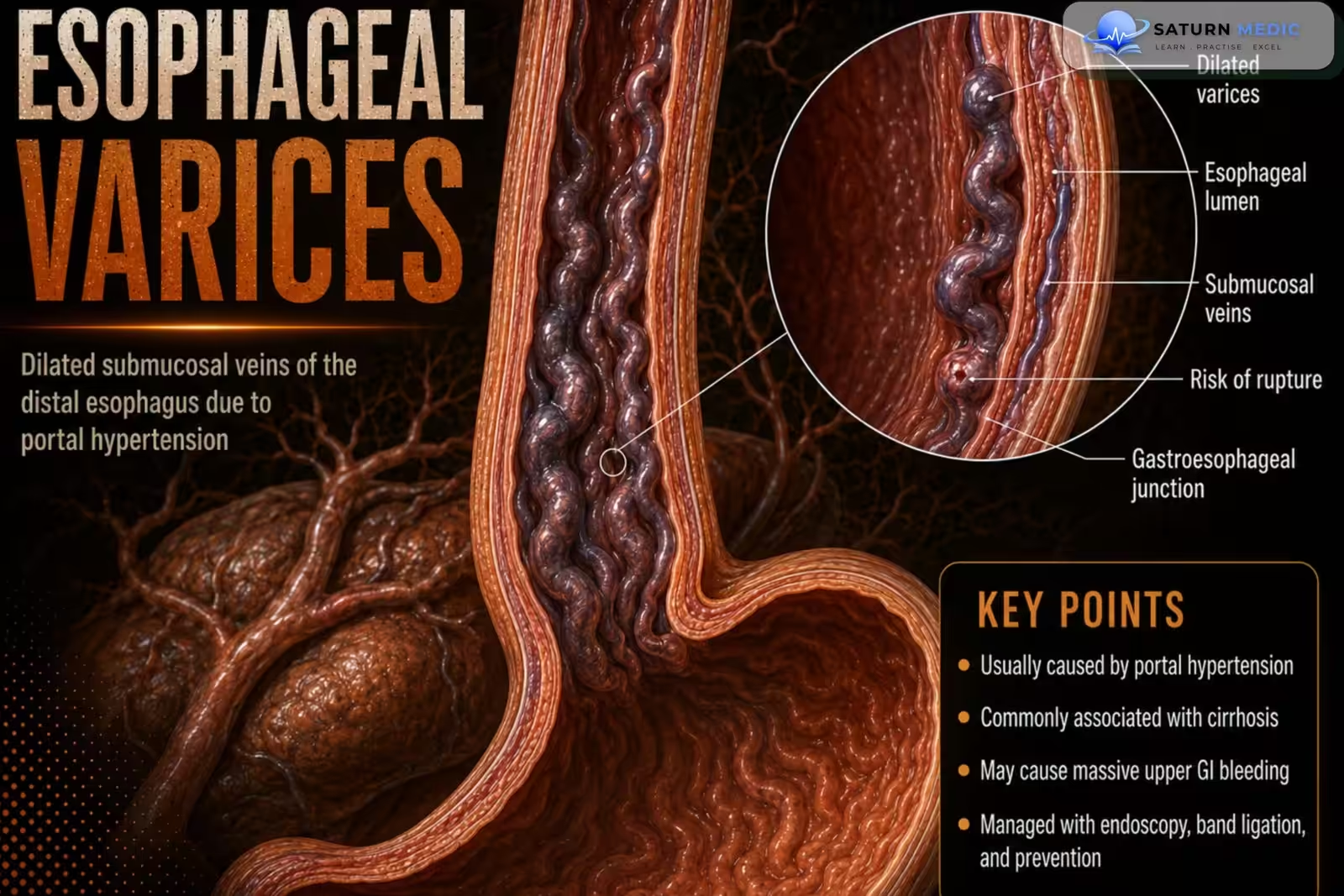
Esophageal varices are dilated collateral veins in the esophagus that develop due to portal
hypertension, most commonly as a result of cirrhosis.
Also called
ICD-10
Specialty
Onset
Reviewed
-
OverviewOverview
-
Etiology & Risk FactorsEtiology & Risk Factors
-
PathophysiologyPathophysiology
-
Clinical PresentationClinical Presentation
-
History TakingHistory Taking
-
Physical ExaminationPhysical Examination
-
InvestigationsInvestigations
-
DiagnosisDiagnosis
-
ManagementManagement
-
ComplicationsComplications
-
PrognosisPrognosis
-
Key Points / Clinical PearlsKey Points / Clinical Pearls
Overview
Esophageal varices form because increased portal venous pressure diverts blood through collateral
veins in the esophagus. Nonbleeding varices are usually asymptomatic. Variceal hemorrhage is a
life-threatening emergency requiring prompt resuscitation, vasoactive therapy, antibiotics, and
endoscopic treatment
Etiology & Risk Factors
Common Causes
- Liver cirrhosis — most common cause
- Chronic viral hepatitis
- Alcohol-related liver disease
- Metabolic dysfunction-associated steatotic liver disease
- Autoimmune or cholestatic liver disease
- Portal vein thrombosis
- Schistosomiasis
- Budd–Chiari syndrome
- Severe right-sided heart failure or constrictive pericarditis
- Congenital hepatic fibrosis
- Splenic vein thrombosis, more commonly causing isolated gastric varices
Portal hypertension caused by cirrhosis, schistosomiasis, or portal vein thrombosis is a major underlying mechanism for variceal formation.
Risk Factors for Developing Varices
- Advanced cirrhosis
- Clinically significant portal hypertension
- Splenomegaly
- Thrombocytopenia
- Decompensated liver disease
- Ascites
- Ongoing alcohol use
- Progressive hepatic fibrosis
Risk Factors for Variceal Bleeding
- Large varices
- High portal pressure
- Red wale marks or red signs on endoscopy
- Advanced liver dysfunction
- Previous variceal hemorrhage
- Active alcohol consumption
- Infection or acute decompensation
- Poor liver reserve
Pathophysiology
1. Portal hypertension increases portal venous pressure.
2. Blood is diverted through collateral venous pathways.
3. Dilated submucosal veins develop in the distal esophagus (esophageal varices).
4. Increased wall tension predisposes the varices to rupture and hemorrhage.
Clinical Presentation
-Nonbleeding varices:
• Usually asymptomatic
-Acute variceal hemorrhage:
• Sudden hematemesis
• Melena
• Features of gastrointestinal bleeding
• Hypovolemic shock (in severe cases)
History Taking
Ask about:
• History of cirrhosis or portal hypertension
• Hematemesis
• Melena
• Previous episodes of variceal bleeding
• Symptoms of chronic liver disease
• Alcohol history
• Previous endoscopic treatment
• Use of nonselective beta blockers
Physical Examination
Assess for:
• Signs of upper gastrointestinal bleeding
• Hemodynamic instability
• Hypovolemic shock
• Clinical features of chronic liver disease and portal hypertension
Investigations
Initial Tests
Complete blood count (CBC)
Liver function tests (LFTs)
Coagulation profile (PT/INR)
Renal function tests and electrolytes
Blood type and crossmatch (if bleeding is suspected)
Diagnostic Investigations
Upper Gastrointestinal Endoscopy (EGD) – Gold Standard
Gold standard for diagnosis
Confirms the presence, size, and location of varices
Identifies high-risk stigmata (e.g., red wale signs, red spots)
Allows therapeutic intervention (e.g., band ligation)
Imaging
Abdominal ultrasound with Doppler
Evaluates liver morphology
Assesses portal vein patency
Detects portal hypertension and splenomegaly
CT or MRI abdomen (when indicated)
Evaluates portal venous system
Assesses complications of portal hypertension
Additional Investigations
Transient elastography (FibroScan) to assess liver fibrosis (selected patients)
Hepatitis B and C serology
Ascitic fluid analysis if ascites is present
Diagnosis
-Diagnosis is established by:
- Esophagogastroduodenoscopy (EGD)
- Patients with cirrhosis should undergo routine screening to detect varices before bleeding occurs
Related Topics
Management
1. Primary Prevention (Non-Bleeding Varices)
Nonselective beta-blockers (NSBBs)
Carvedilol
Propranolol
Nadolol
Endoscopic variceal ligation (EVL) for patients with large varices or those who cannot tolerate beta-blockers.
2. Acute Variceal Bleeding
Initial Stabilization
Airway protection if indicated
Oxygen therapy
Two large-bore IV lines
Fluid resuscitation
Restrictive blood transfusion strategy (target Hb ≈ 7–9 g/dL)
Medical Therapy
Vasoactive drugs
Octreotide
Terlipressin
Prophylactic intravenous antibiotics
e.g., Ceftriaxone
Endoscopic Therapy
Endoscopic variceal ligation (EVL) – First-line treatment
Endoscopic sclerotherapy if band ligation is unavailable or unsuccessful
Rescue Therapy
Balloon tamponade (temporary bridge)
Transjugular intrahepatic portosystemic shunt (TIPS) for uncontrolled or recurrent bleeding
3. Secondary Prevention
After recovery from the first bleed:
Combination of:
Nonselective beta-blocker
Repeated endoscopic variceal ligation
Evaluate for liver transplantation in patients with advanced cirrhosis
Diagnosis is established by:
• Esophagogastroduodenoscopy (EGD)
Patients with cirrhosis should undergo routine screening to detect varices before bleeding occursTreat the underlying cause of portal hypertension
Manage chronic liver disease
Alcohol cessation (if alcohol-related liver disease)
Liver transplantation for end-stage liver disease
Complications
• Variceal hemorrhage
• Hypovolemic shock
• Recurrent bleeding
Prognosis
- The prognosis depends on the severity of the underlying liver disease and whether bleeding occurs.
- Early screening and appropriate primary and secondary prophylaxis reduce the risk of recurrent
hemorrhage.
Key Points / Clinical Pearls
• Esophageal varices are caused by portal hypertension, most commonly due to cirrhosis.
• Nonbleeding varices are usually asymptomatic.
• EGD is recommended for screening in patients with cirrhosis.
• Acute bleeding presents with hematemesis, melena, and may cause hypovolemic shock.
• Acute management includes stabilization, vasoactive drugs, antibiotics, and urgent endoscopic
therapy.
• Persistent bleeding may require balloon tamponade or emergent TIPS.
• Primary prophylaxis includes NSBBs or EVL.
• Secondary prophylaxis includes NSBBs, EVL, and selected use of TIPS.
- Meseeha M, Tian C, Attia M. National Center for Biotechnology Information (NIH). Esophageal Varices, StatPearls.
- Kaplan DE, Ripoll C, Thiele M, et al. AASLD Practice Guidance on Risk Stratification and Management of Portal Hypertension and Varices in Cirrhosis. Hepatology. 2024;79:1180-1211. PMID: 37870298.
- de Franchis R, Bosch J, Garcia-Tsao G, Reiberger T, Ripoll C; Baveno VII Faculty. Baveno VII - Renewing Consensus in Portal Hypertension. J Hepatol. 2022;76:959-974. PMID: 35120736.
- MedlinePlus, National Library of Medicine (NIH). Esophageal Varices: Medical Encyclopedia.
- Singh S, Chandan S, Vinayek R, Aswath G, Facciorusso A, Maida M. Comprehensive Approach to Esophageal Variceal Bleeding: From Prevention to Treatment. World J Gastroenterol. 2024;30:4602-4608. PMID: 39575399.