

Clinical Subject Page
Hepatocellular Carcinoma (HCC)
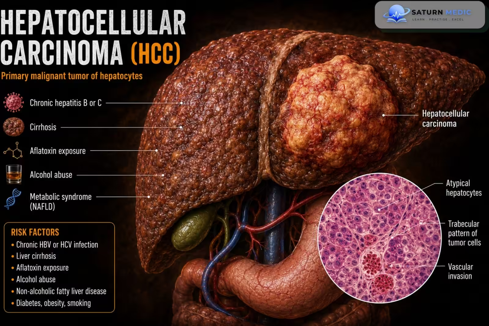
Hepatocellular carcinoma (HCC) is the most common primary malignant tumor of the liver. It usually
develops in patients with chronic liver disease, especially liver cirrhosis or chronic viral hepatitis.
ICD-10
Specialty
Onset
Reviewed
-
OverviewOverview
-
Etiology & Risk FactorsEtiology & Risk Factors
-
PathophysiologyPathophysiology
-
Clinical PresentationClinical Presentation
-
History TakingHistory Taking
-
Physical ExaminationPhysical Examination
-
InvestigationsInvestigations
-
DiagnosisDiagnosis
-
ManagementManagement
-
ComplicationsComplications
-
PrognosisPrognosis
-
Key Points / Clinical PearlsKey Points / Clinical Pearls
Overview
•Hepatocellular Carcinoma (HCC) is the Most common primary liver cancer.
• Usually develops in patients with cirrhosis or chronic hepatitis B/C.
• Early disease is often asymptomatic.
• High-risk patients require surveillance with abdominal ultrasound every 6 months (± AFP).
• Management depends on tumor stage, liver function, and patient performance status.
Etiology & Risk Factors
• Liver cirrhosis (any cause)
• Chronic hepatitis B
• Chronic hepatitis C
• Alcohol-associated liver disease
• Metabolic dysfunction-associated steatotic liver disease (MASLD)
• Hemochromatosis
• Other chronic liver diseases
Pathophysiology
• Chronic liver injury leads to fibrosis and cirrhosis.
• Persistent inflammation promotes malignant transformation of hepatocytes.
• Tumor growth may invade blood vessels (especially the portal vein) and metastasize.
Clinical Presentation
-Early Hepatocellular Carcinoma (HCC) :
• Often asymptomatic.
-Symptomatic disease:
• Right upper quadrant abdominal pain
• Weight loss
• Anorexia
• Fatigue
• Jaundice
• Ascites
• Features of chronic liver disease
• Hepatomegaly
History Taking
-Ask about:
• History of cirrhosis
• Chronic hepatitis B or C
• Alcohol use
• Weight loss
• Anorexia
• Abdominal pain
• Jaundice
• Ascites
• Previous liver lesions
• Family history of liver disease
• Previous HCC surveillance
Physical Examination
• General appearance
• Jaundice
• Hepatomegaly
• Right upper quadrant tenderness
• Ascites
• Splenomegaly
• Peripheral edema
• Signs of chronic liver disease
• Signs of portal hypertension
Investigations
1. Initial investigations for Hepatocellular Carcinoma (HCC)
• Abdominal ultrasound (preferred initial imaging)
• Serum alpha-fetoprotein (AFP) (may improve detection)
2. Diagnostic imaging
If: Liver lesion ≥10 mm, or AFP ≥20 ng/mL
→ Perform multiphase contrast-enhanced CT or MRI.
-Characteristic imaging findings
• Arterial phase hyperenhancement
• Nonperipheral portal venous washout
• Enhancing capsule
• Possible vascular invasion
3. Laboratory studies
Liver assessment
• Liver chemistries
• Coagulation profile
• CBC
• AFP (typically elevated)
Evaluate underlying liver disease
• Hepatitis panel
• Iron studies (if indicated)
4. Liver biopsy
Not routinely required in patients with cirrhosis and characteristic imaging findings.
Indications: Inconclusive multiphase imaging; Suspicious lesion in patients without cirrhosis.
Risks: Bleeding; Tumor seeding.
5. Staging
Following confirmed diagnosis, perform staging investigations to determine disease extent and
guide treatment
Diagnosis
-Diagnosis of Hepatocellular Carcinoma (HCC) is based on:
• Clinical suspicion
• Ultrasound screening
• AFP (adjunctive)
• Characteristic multiphase CT or MRI findings
• Liver biopsy when indicated
• Staging after diagnosis
Related Topics
Management
General Measures for Hepatocellular Carcinoma (HCC)
-
Manage in a multidisciplinary liver cancer team.
-
Treat the underlying liver disease (HBV, HCV, alcohol, NAFLD).
-
Optimize liver function and nutritional status.
-
Assess liver function (Child-Pugh score) and performance status.
Curative Treatment (Early-Stage HCC)
Surgical Resection
-
First-line for patients with:
-
Solitary tumor
-
Preserved liver function
-
No significant portal hypertension
-
Liver Transplantation
-
Best option for selected patients with cirrhosis.
-
Consider patients meeting Milan criteria:
-
Single tumor ≤5 cm, or
-
Up to 3 tumors, each ≤3 cm
-
No vascular invasion or extrahepatic spread
-
Local Ablation
-
Radiofrequency ablation (RFA)
-
Microwave ablation (MWA)
Best for:
-
Small tumors (generally ≤3 cm)
-
Patients unsuitable for surgery
Locoregional Therapy (Intermediate-Stage HCC)
Transarterial Chemoembolization (TACE)
-
Standard treatment for unresectable intermediate-stage HCC.
-
Delivers chemotherapy directly into the hepatic artery followed by embolization.
Transarterial Radioembolization (TARE/Y-90)
-
Alternative to TACE in selected patients.
-
Particularly useful when portal vein thrombosis is present.
Systemic Therapy (Advanced HCC)
First-line options:
-
Atezolizumab + Bevacizumab (preferred)
-
Durvalumab + Tremelimumab (STRIDE regimen)
-
Lenvatinib
-
Sorafenib
Second-line options:
-
Regorafenib
-
Cabozantinib
-
Ramucirumab (AFP ≥400 ng/mL)
-
Immune checkpoint inhibitors in selected patients
Palliative and Supportive Care
-
Pain control
-
Nutritional support
-
Management of ascites
-
Treatment of hepatic encephalopathy
-
Control of variceal bleeding
-
Psychological and palliative care support
Complications
• Portal vein invasion
• Liver failure
• Tumor recurrence
• Metastatic disease
• Death
Prognosis
• prognosis of Hepatocellular Carcinoma (HCC) Depends on tumor stage, liver function, and treatment.
• Early-stage disease may be cured with resection, transplantation, or ablation.
• Advanced disease has a poor prognosis.
• Prognosis is often limited by underlying cirrhosis
Key Points / Clinical Pearls
• Hepatocellular Carcinoma (HCC) is the most common primary liver cancer.
• Most patients have underlying cirrhosis or chronic HBV/HCV.
• Ultrasound is the preferred initial screening test.
• AFP is an adjunctive marker.
• Multiphase CT/MRI confirms diagnosis.
• Classic imaging: arterial enhancement + portal venous washout.
• Biopsy is not routinely required if imaging is characteristic in cirrhotic patients.
• Early disease → resection, transplantation, or radiofrequency ablation.
• Intermediate disease → TACE or TARE.
• Advanced disease → atezolizumab + bevacizumab (first-line systemic therapy).
• End-stage disease → supportive care
- National Center for Biotechnology Information (NIH). Hepatocellular Carcinoma, StatPearls.
- Singal AG, Llovet JM, Yarchoan M, et al. AASLD Practice Guidance on Prevention, Diagnosis, and Treatment of Hepatocellular Carcinoma. Hepatology. 2023;78:1922-1965. Guideline Summary.
- American Association for the Study of Liver Diseases (AASLD). Why Does Treatment of Hepatocellular Carcinoma Require a Multidisciplinary Approach?.
- National Cancer Institute (NIH). Adult Primary Liver Cancer Treatment (PDQ), Health Professional Version.
- MedlinePlus, National Library of Medicine (NIH). Liver Cancer: Medical Encyclopedia.