

Clinical Subject Page
Hypertrophic Pyloric Stenosis
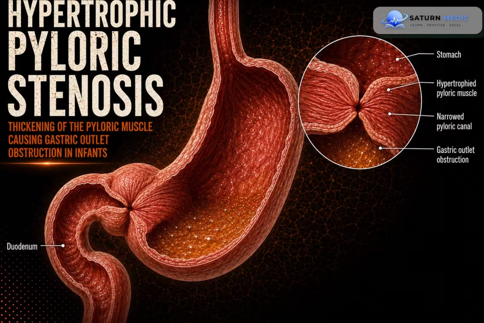
Hypertrophic pyloric stenosis is the most common cause of gastric outlet obstruction in infants,
resulting from hypertrophy of the pyloric muscle that obstructs gastric emptying.
Also called
ICD-10
Specialty
Onset
Reviewed
-
OverviewOverview
-
Etiology & Risk FactorsEtiology & Risk Factors
-
PathophysiologyPathophysiology
-
Clinical PresentationClinical Presentation
-
History TakingHistory Taking
-
Physical ExaminationPhysical Examination
-
InvestigationsInvestigations
-
DiagnosisDiagnosis
-
ManagementManagement
-
ComplicationsComplications
-
PrognosisPrognosis
-
Key Points / Clinical PearlsKey Points / Clinical Pearls
Overview
Hypertrophic pyloric stenosis typically presents between 2 and 6 weeks of age with postprandial
nonbilious projectile vomiting. An olive-shaped mass may be palpable in the epigastrium. The
diagnosis is confirmed with abdominal ultrasound. Before definitive treatment, patients require
rehydration and correction of electrolyte abnormalities, followed by pyloromyotomy.
Etiology & Risk Factors
Common Causes
- Idiopathic hypertrophy of the pyloric muscle (most common)
- Genetic predisposition
- Abnormal innervation of the pylorus (proposed)
- Reduced nitric oxide synthase activity causing impaired pyloric relaxation (proposed)
Risk Factors
- Male sex (especially first-born males)
- Family history of hypertrophic pyloric stenosis
- Age 2–8 weeks (most commonly 3–6 weeks)
- Macrolide antibiotic exposure (especially erythromycin or azithromycin in early infancy)
- Formula feeding
- Maternal smoking during pregnancy
- Prematurity (less common)
Pathophysiology
1. Hypertrophy of the pyloric muscle develops.
2. The pyloric canal progressively narrows.
3. Gastric emptying becomes obstructed.
4. Infants develop forceful nonbilious vomiting after feeding.
5. Persistent vomiting leads to dehydration and electrolyte disturbances
Clinical Presentation
Typical features include:
• Postprandial nonbilious projectile vomiting
• Symptom onset between 2 and 6 weeks of age
• Palpable olive-shaped epigastric mass
History Taking
-
Ask about:
• Age at symptom onset (typically 2–6 weeks)
• Projectile vomiting after feeds
• Frequency and duration of vomiting
• Feeding history
• Decreased urine output or other signs of dehydration
• Weight gain or weight loss
Physical Examination
Assess for:
• Olive-shaped mass in the epigastrium
• Signs of dehydration
• Nutritional status
Investigations
Initial Tests
-
Complete blood count (CBC)
-
Electrolytes
-
Renal function tests
-
Blood gas analysis (if severe vomiting)
Typical findings:
-
Hypochloremic, hypokalemic metabolic alkalosis
-
Dehydration
Diagnostic Investigations
Abdominal Ultrasound – Gold Standard
-
Investigation of choice
-
Demonstrates:
-
Thickened pyloric muscle
-
Elongated pyloric canal
-
Failure of gastric contents to pass through the pylorus
-
Upper Gastrointestinal Contrast Study
Performed if ultrasound is inconclusive.
May show:
-
String sign
-
Delayed gastric emptying
-
Narrow pyloric canal
Additional Investigations
-
Assess hydration status
-
Monitor urine output before surgery
Diagnosis
-Diagnosis is established by:
• Typical clinical presentation
• Confirmation with abdominal ultrasound
Related Topics
Management
1. Conservative Management
For small, asymptomatic diverticula:
Observation
Regular follow-up
Dietary modification (eat slowly, chew food thoroughly, drink water with meals)
2. Endoscopic Treatment (Preferred)
First-line treatment for most symptomatic patients.
Options include:
Endoscopic diverticulotomy (septotomy)
Endoscopic stapling diverticulotomy
Flexible endoscopic septal division (FESD)
Zenker’s peroral endoscopic myotomy (Z-POEM) (selected patients)
These procedures divide the septum between the diverticulum and the esophagus, improving esophageal emptying.
3. Surgical Treatment
Indicated for:
Large diverticula
Failed endoscopic treatment
Recurrence after endoscopic therapy
Patients unsuitable for endoscopic procedures
Options include:
Diverticulectomy with cricopharyngeal myotomy
Diverticulopexy with cricopharyngeal myotomy
Diverticular inversion (rarely)
4. Supportive Care
Nutritional assessment
Treat aspiration pneumonia if present
Manage dehydration or malnutrition
Speech and swallowing therapy when indicated
5. Follow-Up
Monitor for symptom recurrence
Assess swallowing function
Repeat endoscopy or barium swallow if symptoms recur
Complications
• Gastric outlet obstruction
• Dehydration
• Electrolyte abnormalities
Prognosis
- The prognosis is excellent with prompt diagnosis, correction of fluid and electrolyte disturbances,
and definitive treatment by pyloromyotomy
Key Points / Clinical Pearls
• Hypertrophic pyloric stenosis is the most common cause of gastric outlet obstruction in infants.
• Symptoms usually begin between 2 and 6 weeks of age.
• Projectile nonbilious vomiting is the hallmark symptom.
• An olive-shaped epigastric mass is a classic examination finding.
• Abdominal ultrasound is the diagnostic test of choice.
• Correct dehydration and electrolyte abnormalities before surgery.
• Pyloromyotomy is the definitive treatment.
- National Center for Biotechnology Information (NIH). Pyloric Stenosis, StatPearls.
- Infantile Hypertrophic Pyloric Stenosis at a Tertiary Care Hospital in Tanzania: A Surgical Experience With 102 Patients Over a 5-Year Period. PMC4652415.
- MedlinePlus, National Library of Medicine (NIH). Pyloric Stenosis: Medical Encyclopedia.
- Hypertrophic Pyloric Stenosis in a Four-Week-Old Infant: Radiological Diagnosis and Pitfalls. PMC12776224.
- Hulka F, Harrison MW, Campbell TJ, Campbell JR. Complications of Pyloromyotomy for Infantile Hypertrophic Pyloric Stenosis. Am J Surg. 1997;173:450-452. PMID: 9168087.