

Clinical Subject Page
Mesenteric Ischemia
Zenker diverticulum is the most common upper esophageal (pharyngoesophageal) diverticulum. It
is a false (pulsion) diverticulum that arises through the Killian triangle, a weak area in the posterior
wall of the hypopharynx.
Also called
ICD-10
Specialty
Onset
Reviewed
-
OverviewOverview
-
Etiology & Risk FactorsEtiology & Risk Factors
-
PathophysiologyPathophysiology
-
Clinical PresentationClinical Presentation
-
History TakingHistory Taking
-
Physical ExaminationPhysical Examination
-
InvestigationsInvestigations
-
DiagnosisDiagnosis
-
ManagementManagement
-
ComplicationsComplications
-
PrognosisPrognosis
-
Key Points / Clinical PearlsKey Points / Clinical Pearls
Overview
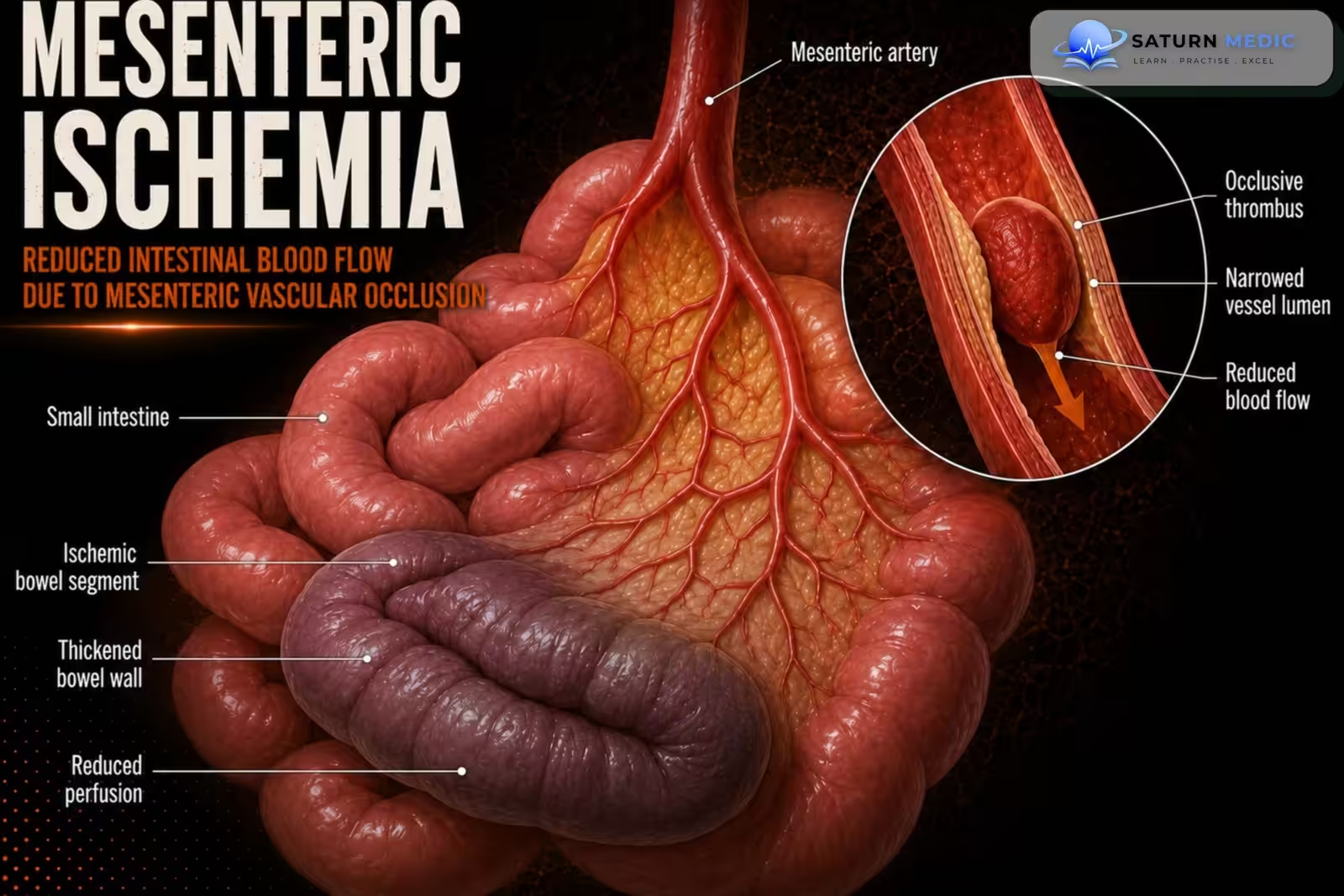
Mesenteric Ischemia is a life-threatening reduction in blood flow to the intestines, leading to bowel ischemia and possible necrosis. It most commonly results from arterial embolism, arterial thrombosis, mesenteric venous thrombosis, or nonocclusive hypoperfusion. Patients typically present with sudden severe abdominal pain that is disproportionate to physical examination findings. Early diagnosis with CT angiography and prompt revascularization are essential to reduce morbidity and mortality.
Etiology & Risk Factors
-Etiology
- Arterial embolism (50%) – Most common cause
- Usually affects the superior mesenteric artery (SMA)
- Arterial thrombosis (25%)
- Due to atherosclerosis
- Nonocclusive mesenteric ischemia (20%)
- Due to low blood flow (no vessel blockage)
- Mesenteric venous thrombosis (<10%)
- Least common cause
-Risk Factors
- Atrial fibrillation (most important risk factor for embolism)
- Atherosclerosis (most important risk factor for thrombosis)
- Hypotension/low cardiac output (critically ill patients)
- Vasopressor use
- Hypercoagulable states
- Malignancy
- Portal hypertension
- Estrogen therapy
Pathophysiology
Mesenteric blood flow (usually SMA occlusion) → ↓ Intestinal oxygen supply (hypoxia) → Intestinal ischemia → Bowel infarction/necrosis → Breakdown of intestinal mucosal barrier → Bacterial & toxin translocation → Perforation → Sepsis → Multiple organ failure → Death (if untreated)
Clinical Presentation
- Sudden, severe abdominal pain that is out of proportion to physical examination (classic finding)
- Nausea and vomiting
- Abdominal bloating
- Diarrhea, becoming bloody in later stages
- Signs of sepsis (fever, tachycardia, hypotension) as the disease progresses
- Peritonitis/acute abdomen in late stages (suggests bowel infarction)
Classic Triad
- Severe abdominal pain
- Bloody diarrhea
- Atrial fibrillation
History Taking
-
Ask about:
- When did the abdominal pain start? (sudden or gradual?)
- Where is the pain?
- How severe is the pain (0–10)?
- Is the pain constant or intermittent?
- Do you have nausea or vomiting?
- Have you had diarrhea or blood in your stool?
- Have you had similar episodes before?
- Do you have atrial fibrillation or heart disease?
- Have you had a heart attack, stroke, or vascular disease?
- Have you recently had low blood pressure, shock, or been critically ill?
- Do you take blood thinners or estrogen therapy?
- Do you have cancer or a blood-clotting disorder?
- Do you use cocaine or vasoconstrictive drugs?
Physical Examination
-
Early Findings
- Severe abdominal pain with minimal abdominal tenderness (pain out of proportion to exam)
- Abdomen may be soft with few physical findings
- Mild abdominal distension (bloating)
Late Findings
- Diffuse abdominal tenderness
- Rebound tenderness
- Guarding/rigidity (peritonitis)
- Absent bowel sounds (may occur with advanced disease)
- Signs of sepsis: fever, tachycardia, hypotension
- Hemodynamic instability/shock (severe disease)
Investigations
-
1. Imaging (Most Important)
- CT angiography (CTA) abdomen and pelvis Test of choice
- Confirms the diagnosis
- Identifies embolism, thrombosis, bowel ischemia/infarction
- Do not delay CTA while waiting for laboratory results.
2. Laboratory Tests
- CBC: leukocytosis
- Serum lactate: increased (poor prognostic sign; may be normal early)
- ABG/VBG: metabolic acidosis, low bicarbonate
- CMP: electrolyte abnormalities, increased AST
- D-dimer: may be elevated (not specific)
- Amylase, CPK, LDH: may be elevated
3. Other Imaging
- Catheter angiography: if endovascular treatment is planned or in selected cases
- Abdominal X-ray: mainly to assess for perforation or obstruction (may be normal)
- CT angiography (CTA) abdomen and pelvis Test of choice
Diagnosis
- Sudden severe abdominal pain out of proportion to the physical exam
- Atrial fibrillation or other vascular risk factors
- Bloody diarrhea (late)
- Signs of peritonitis or sepsis (late)
Confirmatory Test
- CT angiography (CTA) of the abdomen and pelvis = Test of choice
- Detects embolism, thrombosis, bowel ischemia/infarction
- Do not delay CTA if AMI is suspected
Laboratory Findings (Supportive, Not Diagnostic)
- ↑ WBC
- ↑ Lactate (may be normal early)
- Metabolic acidosis
- ↑ D-dimer
Related Topics
Management
-
Initial Management
- ABCDE approach and stabilize the patient
- High-flow oxygen
- Two large-bore IV lines and aggressive IV fluids
- Keep patient NPO
- Insert a nasogastric (NG) tube
- Broad-spectrum IV antibiotics
- IV unfractionated heparin (if no contraindication and coordinated with surgery)
- Adequate analgesia and antiemetics
- Urgent surgical and vascular/interventional radiology consultation
- Admit to ICU
Definitive Treatment
- Endovascular revascularization (embolectomy, thrombectomy, angioplasty ± stent) in appropriate stable patients
Emergency laparotomy if there is:
- Peritonitis
- Bowel infarction/necrosis
- Hemodynamic instability
- Bowel resection if necrotic bowel is present
Complications
- Bowel infarction (intestinal necrosis)
- Peritonitis
- Sepsis
- Multiple organ failure (MOF)
- Abdominal compartment syndrome
- Death (if diagnosis and treatment are delayed)
- Peritonitis indicates bowel infarction and requires emergency surgery.
- Delayed diagnosis greatly increases the risk of sepsis, multiple organ failure, and death.
Prognosis
- Poor prognosis if diagnosis or treatment is delayed
- Overall mortality: 50–70%
- Early treatment (within 6 hours) can reduce mortality to 10–20%
- Prognosis is worse if there is:
- Bowel infarction (necrosis)
- Perforation
- Sepsis
- Multiple organ failure
Key Points / Clinical Pearls
- Vascular emergency with high mortality (50–70%).
- Most common cause: Superior mesenteric artery (SMA) embolism.
- Most important risk factor: Atrial fibrillation.
- Hallmark symptom: Severe abdominal pain out of proportion to the physical examination.
- Classic triad: Severe abdominal pain + bloody diarrhea + atrial fibrillation.
- CT angiography (CTA) is the test of choice.
- Do not delay CTA while waiting for laboratory results.
- Normal lactate does not exclude acute mesenteric ischemia early in the disease.
- Initial treatment: ABCDE, oxygen, IV fluids, NPO, NG tube, IV antibiotics, IV heparin (if appropriate), urgent surgical/vascular consultation.
- Peritonitis or hemodynamic instability = emergency laparotomy
- Zafer S, Lopez RA, Kimyaghalam A. National Center for Biotechnology Information (NIH). Acute Mesenteric Ischemia, StatPearls.
- Björck M, Koelemay M, Acosta S, et al; ESVS Guidelines Committee. Management of the Diseases of Mesenteric Arteries and Veins: Clinical Practice Guidelines of the European Society of Vascular Surgery (ESVS). Eur J Vasc Endovasc Surg. 2017;53:460-510. ESVS Guideline.
- Bala M, Kashuk J, Moore EE, et al. Acute Mesenteric Ischemia: Guidelines of the World Society of Emergency Surgery. World J Emerg Surg. 2017;12:38. PMC5545843.
- MedlinePlus, National Library of Medicine (NIH). Mesenteric Artery Ischemia: Medical Encyclopedia.
- National Center for Biotechnology Information (NIH). Bowel Ischemia, StatPearls.