

Clinical Subject Page
Pancreatic Cancer
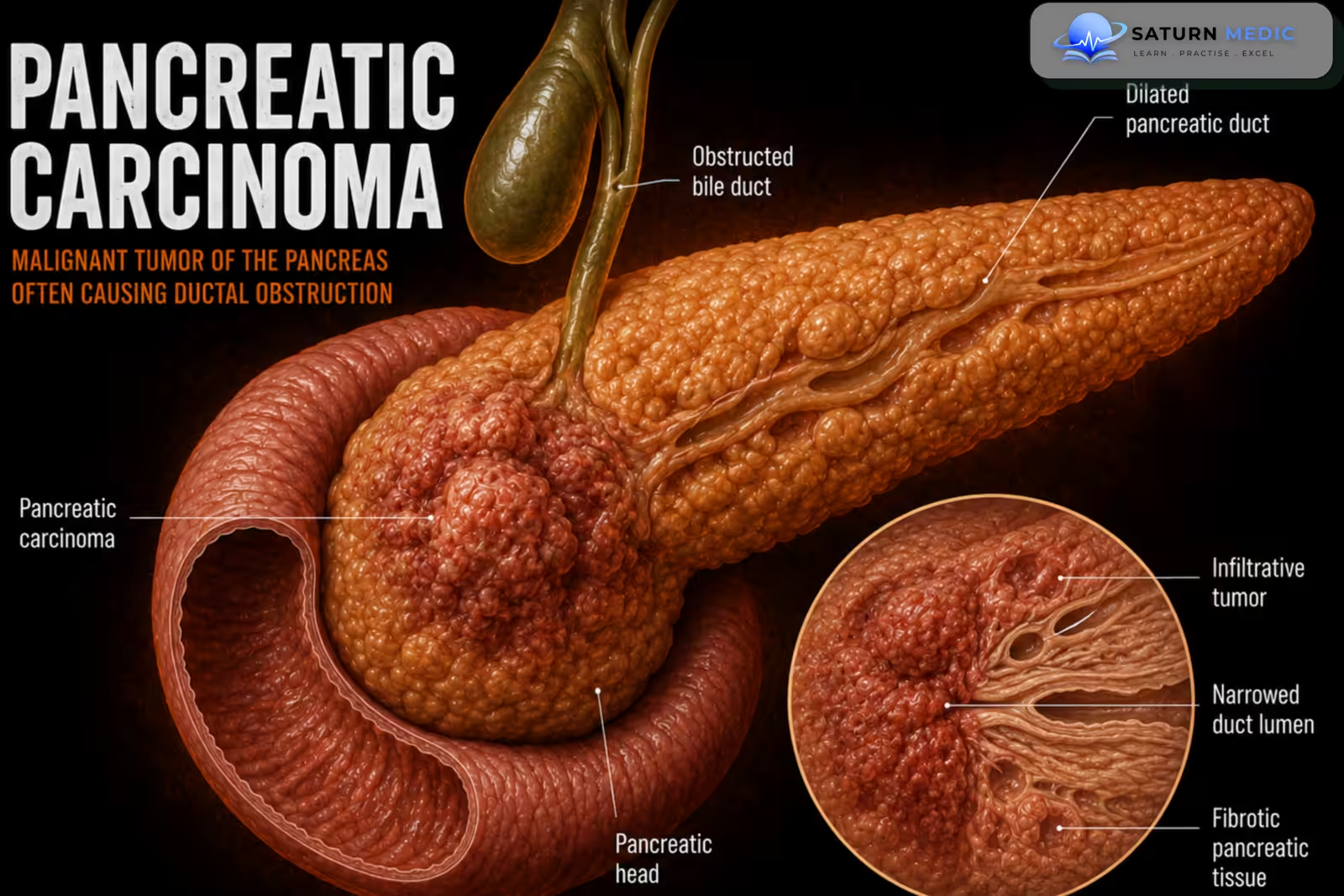
Pancreatic carcinoma is a malignant (cancerous) tumor of the pancreas. Most pancreatic cancers are ductal adenocarcinomas, which arise from the pancreatic ducts. It is an aggressive cancer that is often diagnosed late because symptoms usually appear only after the disease has advanced.
Also called
ICD-10
Specialty
Onset
Reviewed
-
OverviewOverview
-
Etiology & Risk FactorsEtiology & Risk Factors
-
PathophysiologyPathophysiology
-
Clinical PresentationClinical Presentation
-
History TakingHistory Taking
-
Physical ExaminationPhysical Examination
-
InvestigationsInvestigations
-
DiagnosisDiagnosis
-
ManagementManagement
-
ComplicationsComplications
-
PrognosisPrognosis
-
Key Points / Clinical PearlsKey Points / Clinical Pearls
Overview
- Usually affects adults 60–80 years of age.
- Common risk factors include smoking, obesity, heavy alcohol consumption, and chronic pancreatitis.
- Most tumors develop in the head of the pancreas.
Etiology & Risk Factors
-Etiology and Risk factors for Pancreatic Cancer
- The exact cause of pancreatic carcinoma is unknown. It develops due to genetic changes that cause uncontrolled growth of pancreatic cells.
- Risk Factors
- Smoking (strongest risk factor)
- Age > 50 years
- Chronic pancreatitis (especially long-standing)
- Obesity
- Type 2 diabetes mellitus
- High alcohol consumption
- Occupational exposure to chemicals (e.g., dry cleaning and metalworking industries)
- Inherited genetic syndromes/family history (about 10% of cases), including BRCA mutations, Peutz-Jeghers syndrome, hereditary pancreatitis, and familial pancreatic cancer
Pathophysiology
Risk factors/genetic mutations → Uncontrolled growth of pancreatic ductal epithelial cells → Ductal adenocarcinoma develops → Tumor enlarges (usually in the head of the pancreas) → Bile duct and pancreatic duct obstruction → Painless jaundice ± pancreatitis → Local invasion of nearby tissues → Spread (metastasis), commonly to the liver → Weight loss, pain, advanced disease.
Clinical Presentation
Early stage of Pancreatic Cancer
- Usually no symptoms
Constitutional symptoms
- Weight loss
- Poor appetite
- Weakness
Gastrointestinal symptoms
- Epigastric pain radiating to the back
- Nausea
- Painless jaundice (especially with tumors in the head of the pancreas)
- Pale stools
- Dark urine
- Itching (pruritus)
- Diarrhea/steatorrhea (fatty stools) due to malabsorption
Other features of Pancreatic Cancer
- Hypercoagulability (e.g., Trousseau syndrome with migratory thrombophlebitis)
History Taking
Ask about:
- When did your symptoms start?
- Do you have upper abdominal (epigastric) pain? Does it radiate to your back?
- Have you noticed yellowing of your eyes or skin (jaundice)?
- Is your urine dark or your stool pale?
- Do you have itching (pruritus)?
- Have you had unintentional weight loss?
- Have you lost your appetite?
- Do you have nausea or vomiting?
- Have you noticed fatty stools (steatorrhea) or diarrhea?
- Have you recently developed diabetes or worsening blood sugar control?
- Do you smoke or have you smoked in the past?
- How much alcohol do you drink?
- Have you ever had chronic pancreatitis?
- Do you have a family history of pancreatic cancer or inherited cancer syndromes (e.g., BRCA mutation)?
- Have you had any previous pancreatic or biliary disease?
Physical Examination
General Examination of Pancreatic Cancer
- Weight loss and cachexia
- Weakness and fatigue
- Jaundice (yellow skin and sclera)
- Scratch marks due to itching (pruritus)
Vital Signs
- Usually normal in early disease
- Fever may be present if cholangitis or infection develops
Abdominal Examination
- Epigastric or right upper quadrant tenderness
- Palpable nontender enlarged gallbladder (Courvoisier sign) with painless obstructive jaundice
- Palpable abdominal mass (occasionally)
- Hepatomegaly if liver metastases are present
- Ascites in advanced disease
Extremities
- Signs of deep vein thrombosis (DVT)
- Migratory superficial thrombophlebitis (Trousseau syndrome)
Investigations
1. Laboratory Tests
- CBC – anemia
- Liver function tests (LFTs) – ↑ bilirubin, ALP, GGT (obstructive jaundice)
- Amylase and lipase
- Blood glucose – may be elevated
- CA 19-9 – tumor marker (used for prognosis and monitoring, not for screening or diagnosis)
- CEA – less specific, may be used as an adjunct
2. Imaging
- Abdominal ultrasound – first test, especially if jaundice is present
- Contrast-enhanced CT abdomen (pancreas protocol) – investigation of choice for detecting the tumor, assessing spread, and determining resectability
- MRI/MRCP – if CT is contraindicated or for better evaluation of bile and pancreatic ducts
3. Confirmatory Tests
- Endoscopic ultrasound (EUS) with fine-needle aspiration (FNA) – gold standard for tissue diagnosis
- Biopsy – confirms pancreatic carcinoma
4. Additional Tests
- ERCP – if biliary obstruction is present; allows stent placement and tissue sampling
- CT chest (or CXR) – to assess for metastasis (staging)
Diagnosis
-Diagnosis of Pancreatic Cancer is based on :
- Clinical suspicion: Painless jaundice, weight loss, epigastric pain radiating to the back
- Abdominal ultrasound: Initial test, especially in patients with jaundice
- Contrast-enhanced CT abdomen (pancreas protocol): Best imaging test to detect the tumor, assess local invasion, metastasis, and resectability
- MRI/MRCP: Alternative if CT is contraindicated or for further evaluation of the pancreatic and biliary ducts
- Endoscopic ultrasound (EUS) with fine-needle aspiration (FNA): Confirms the diagnosis by obtaining tissue for histopathology
- Tumor markers: CA 19-9 (and sometimes CEA) are used for prognosis and monitoring, not for diagnosis or screening
- CT chest (or CXR): Used for staging to assess distant metastasis
Related Topics
Management
1. Resectable Pancreatic Cancer (Curative)
- Surgical resection (only curative treatment)
- Whipple procedure for tumors in the head of the pancreas
- Distal pancreatectomy ± splenectomy for body/tail tumors
- Adjuvant chemotherapy after surgery
- Neoadjuvant therapy may be used for borderline resectable tumors before surgery
2. Unresectable or Metastatic Pancreatic Carcinoma (Palliative)
- Combination chemotherapy (e.g., FOLFIRINOX or gemcitabine-based regimens)
- Radiotherapy in selected patients
3. Supportive (Palliative) Care
- Pain control (WHO analgesic ladder; consider celiac plexus block if severe pain)
- Nutritional support and pancreatic enzyme replacement if needed
- Treat biliary obstruction with ERCP and biliary stent (preferred)
- Manage complications and provide palliative care
Complications
- Metastasis (most commonly to the liver, lymph nodes, lungs, and adjacent organs)
- Obstructive jaundice due to bile duct obstruction
- Cholangitis (infection of the bile ducts)
- Duodenal/gastric outlet obstruction
- Ascites (malignant ascites)
- Venous thromboembolism (VTE), including DVT and pulmonary embolism
- Migratory thrombophlebitis (Trousseau syndrome)
- Disseminated intravascular coagulation (DIC)
- Secondary diabetes mellitus
- Malnutrition and weight loss (cachexia)
- The most common cause of death in pancreatic carcinoma is metastatic disease, especially liver metastasis.
Prognosis
- Pancreatic Cancer has a Very poor prognosis because it is usually diagnosed at an advanced stage.
- Highly aggressive cancer with early local invasion and metastasis.
- Overall 5-year survival rate: ~12.8% (depends mainly on disease stage).
- Metastatic pancreatic cancer: ~3% 5-year survival.
- Patients who undergo successful surgical resection have a median survival of ~18 months and a 5-year survival of ~20%.
Key Points / Clinical Pearls
- 95% of Pancreatic Cancer are ductal adenocarcinomas.
- Most tumors occur in the head of the pancreas (65%).
- Smoking is the most important risk factor.
- Classic triad: Painless jaundice + weight loss + epigastric pain radiating to the back.
- CT pancreas protocol is the imaging test of choice.
- EUS-guided FNA provides the definitive diagnosis.
- CA 19-9 is used for monitoring, not for screening.
- Whipple procedure is the only potentially curative treatment for resectable head tumors.
- Liver is the most common site of metastasis.
- Prognosis is poor because most patients present with advanced disease.
- National Center for Biotechnology Information (NIH). Pancreatic Pseudocyst, StatPearls.
- Banks PA, Bollen TL, Dervenis C, et al; Acute Pancreatitis Classification Working Group. Classification of Acute Pancreatitis - 2012: Revision of the Atlanta Classification and Definitions by International Consensus. Gut. 2013;62:102-111.
- Pancreatic Pseudocyst: The Past, the Present, and the Future. World J Gastroenterol. PMC11287700.
- Systematic Review Comparing Endoscopic, Percutaneous and Surgical Pancreatic Pseudocyst Drainage. World J Gastroenterol. PMC4804189.
- MedlinePlus, National Library of Medicine (NIH). Pancreatic Pseudocyst: Medical Encyclopedia.