

Clinical Subject Page
Sigmoid Volvulus
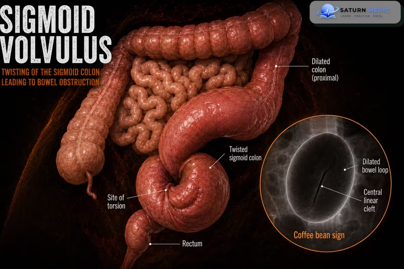
Sigmoid volvulus is a twisting of the sigmoid colon around its mesenteric attachment, producing a closed-loop large-bowel obstruction. Continued twisting may compromise blood flow and cause bowel ischemia, necrosis, perforation, and peritonitis.
Also called
ICD-10
Specialty
Onset
Reviewed
-
OverviewOverview
-
Etiology & Risk FactorsEtiology & Risk Factors
-
PathophysiologyPathophysiology
-
Clinical PresentationClinical Presentation
-
History TakingHistory Taking
-
Physical ExaminationPhysical Examination
-
InvestigationsInvestigations
-
DiagnosisDiagnosis
-
ManagementManagement
-
ComplicationsComplications
-
PrognosisPrognosis
-
Key Points / Clinical PearlsKey Points / Clinical Pearls
Overview
Sigmoid volvulus occurs when a long, mobile sigmoid colon twists around the base of its mesentery. This obstructs the bowel lumen and may also compress the mesenteric blood vessels. Patients commonly present with marked abdominal distension, colicky abdominal pain, constipation or obstipation, nausea, and vomiting. Diagnosis is usually made by abdominal radiography or CT. In patients without ischemia or perforation, urgent endoscopic detorsion is the initial treatment, followed by definitive sigmoid colectomy because recurrence is common
Etiology & Risk Factors
Etiology
Sigmoid volvulus develops when an elongated and redundant sigmoid colon with a narrow mesenteric base rotates around its mesenteric axis.
Risk Factors
Advanced age
Chronic constipation
Redundant or elongated sigmoid colon
Institutionalization
Reduced mobility or prolonged bed rest
Neurological disease
Parkinson disease
Previous stroke
Dementia
Psychiatric illness
Use of medications that reduce bowel motility
High-fiber bulky diet in some populations
Previous abdominal surgery
Chagas disease
Megacolon
Pregnancy, rarely
Pathophysiology
A long, mobile sigmoid colon twists around its narrow mesenteric base, causing obstruction at both ends of the affected segment. Gas and fecal material accumulate within the closed loop, producing progressive colonic distension. Increasing pressure compresses venous drainage first, causing congestion and bowel-wall edema, followed by reduced arterial blood flow. Untreated volvulus can therefore progress to ischemia, gangrene, perforation, fecal peritonitis, sepsis, and shock
Clinical Presentation
Symptoms
Progressive abdominal distension
Colicky or constant abdominal pain
Absolute constipation or obstipation
Inability to pass flatus
Nausea
Vomiting, often occurring late
Reduced appetite
Signs
Markedly distended abdomen
Tympanic percussion note
Abdominal tenderness
Increased bowel sounds early
Reduced or absent bowel sounds late
Empty rectum on digital rectal examination
Tachycardia
Fever
Guarding or rebound tenderness in complicated disease
History Taking
Ask about:
- Onset and progression of abdominal distension
- Abdominal pain
- Last bowel movement
- Ability to pass flatus
- Nausea or vomiting
- Previous similar episodes
- Chronic constipation
- Neurological or psychiatric disease
- Reduced mobility or institutionalization
- Previous abdominal surgery
- Medications that reduce bowel motility
- Fever or rectal bleeding
Physical Examination
General Examination
Assess for:
Fever
Tachycardia
Hypotension
Dehydration
Confusion
Signs of sepsis or shock
Abdominal Examination
Look for:
Marked generalized distension
Visible bowel loops
Surgical scars
Assess for:
Tympany
Tenderness
Guarding
Rebound tenderness
Bowel sounds
Features of peritonitis
Digital Rectal Examination
May show:
Empty rectum
Absence of stool
Blood, which may suggest mucosal ischemia
Investigations
Laboratory Tests
Complete blood count
C-reactive protein
Renal function and electrolytes
Serum lactate
Arterial or venous blood gas
Liver function tests
Coagulation profile
Blood group and crossmatch
Blood cultures if sepsis is suspected
Abdominal Radiograph
Typical findings include:
Coffee-bean sign
Large dilated sigmoid loop
Bent inner-tube appearance
Apex of the dilated loop directed toward the upper abdomen
Absence of rectal gas
CT Abdomen and Pelvis
CT can demonstrate:
Twisted sigmoid colon
Whirl sign of the twisted mesentery
Bird-beak tapering at the point of torsion
Proximal bowel dilatation
Bowel-wall ischemia
Pneumatosis
Free air or perforation
Alternative causes of obstruction
Flexible Sigmoidoscopy
Flexible sigmoidoscopy can:
Confirm the diagnosis
Assess mucosal viability
Detorse and decompress the colon
Allow placement of a rectal decompression tube
Diagnosis
Diagnosis is based on:
- Typical symptoms of large-bowel obstruction
- Marked abdominal distension
- Characteristic abdominal radiograph or CT findings
- Endoscopic confirmation in suitable patients
- Assessment for bowel ischemia, necrosis, or perforation
Related Topics
Management
Initial Management
Keep the patient nil by mouth
Establish intravenous access
Give intravenous fluids
Correct electrolyte abnormalities
Provide analgesia and antiemetics
Insert a nasogastric tube if significant vomiting is present
Give venous thromboembolism prophylaxis when appropriate
Administer broad-spectrum antibiotics if ischemia, perforation, or sepsis is suspected
Arrange urgent surgical review
Stable Patient Without Ischemia or Perforation
Urgent flexible sigmoidoscopy or colonoscopy should be performed for:
Endoscopic detorsion
Colonic decompression
Assessment of mucosal viability
Placement of a decompression tube
Successful endoscopic decompression should usually be followed by sigmoid colectomy during the same admission or soon afterward, because recurrence after decompression alone is common.
Emergency Surgery
Immediate surgery is indicated for:
Peritonitis
Perforation
Bowel ischemia or gangrene
Hemodynamic instability
Failed endoscopic detorsion
Recurrent volvulus when endoscopic treatment is unsuitable
Surgical options include:
Sigmoid colectomy with primary anastomosis in selected stable patients
Sigmoid colectomy with end colostomy, such as a Hartmann procedure, in unstable patients or when the bowel is gangrenous or perforated
Complications
- Recurrent sigmoid volvulus
- Bowel ischemia
- Colonic gangrene
- Perforation
- Fecal peritonitis
- Sepsis
- Septic shock
- Electrolyte abnormalities
- Acute kidney injury
- Death
Prognosis
- The prognosis is generally good when sigmoid volvulus is diagnosed early and successfully treated before ischemia develops. Delayed presentation, bowel gangrene, perforation, advanced age, and severe comorbidities significantly increase morbidity and mortality. Recurrence is common after endoscopic decompression without definitive surgery.
Key Points / Clinical Pearls
- Sigmoid volvulus is twisting of the sigmoid colon around its mesentery.
- It causes closed-loop large-bowel obstruction.
- Abdominal distension, pain, and obstipation are typical.
- The coffee-bean sign may be visible on abdominal radiography.
- CT may show a whirl sign or bird-beak appearance.
- Endoscopic detorsion is first-line when there is no ischemia or perforation.
- Peritonitis, gangrene, perforation, or failed decompression requires emergency surgery.
- Definitive sigmoid colectomy is recommended because recurrence after decompression alone is common.
- Lieske B, Antunes C. National Center for Biotechnology Information (NIH). Sigmoid Volvulus, StatPearls.
- Le CK, Anand S, Cooper W. National Center for Biotechnology Information (NIH). Volvulus, StatPearls.
- Salati U, McNeill G, Torreggiani WC. The Coffee Bean Sign in Sigmoid Volvulus. Radiology. 2011;258:651-652. PMID: 21273530.
- Fu CG. Appropriate Treatment of Acute Sigmoid Volvulus in the Emergency Setting. World J Gastroenterol. 2013;19:4979-4983.
- MedlinePlus, National Library of Medicine (NIH). Intestinal Obstruction: Medical Encyclopedia.