

Clinical Subject Page
Splenic Rupture
Splenic rupture is a tear or disruption of the spleen that causes internal bleeding into the abdominal cavity. It most commonly follows blunt abdominal trauma and can rapidly become life-threatening
Also called
Splenic laceration
ICD-10
S36.0
Specialty
Gastroenterology
Onset
Acute
Reviewed
July 2026
On This Page
-
OverviewOverview
-
Etiology & Risk FactorsEtiology & Risk Factors
-
PathophysiologyPathophysiology
-
Clinical PresentationClinical Presentation
-
History TakingHistory Taking
-
Physical ExaminationPhysical Examination
-
InvestigationsInvestigations
-
DiagnosisDiagnosis
-
ManagementManagement
-
ComplicationsComplications
-
PrognosisPrognosis
-
Key Points / Clinical PearlsKey Points / Clinical Pearls
Overview
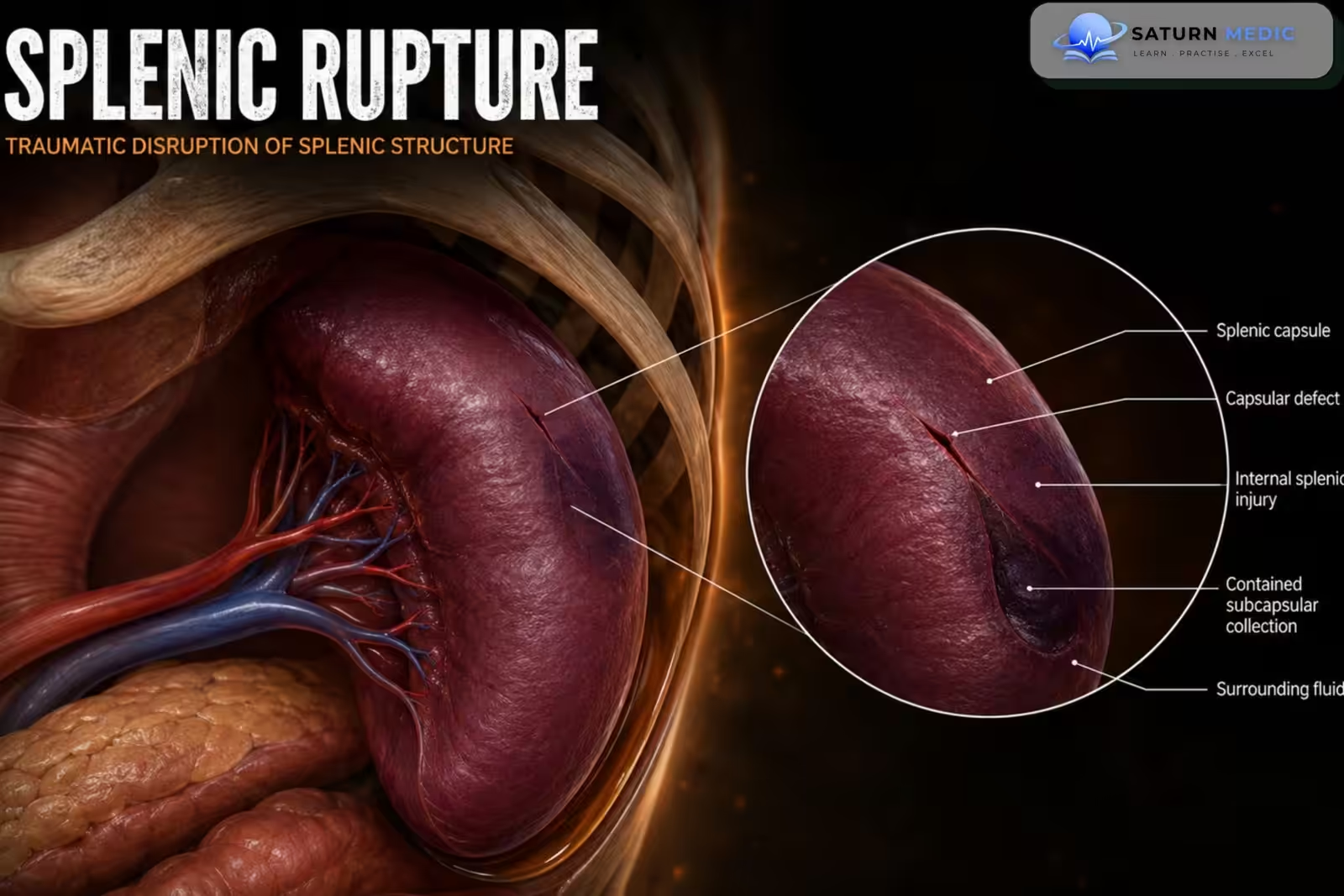
- Splenic rupture occurs when the splenic capsule or tissue is damaged, allowing blood to leak into the peritoneal cavity. The bleeding may occur immediately after injury or may be delayed because of rupture of a subcapsular hematoma. Patients commonly present with left upper-quadrant pain, left shoulder pain, abdominal tenderness, tachycardia, hypotension, or hemorrhagic shock
Etiology & Risk Factors
Traumatic Causes Causing Splenic Rupture
Road traffic accidents
Falls from height
Sports injuries
Direct blow to the left upper abdomen
Fractured left lower ribs
Penetrating abdominal injury
Crushing injury
Iatrogenic injury during abdominal surgery
Blunt abdominal trauma is the most common cause of splenic rupture.
Atraumatic Causes Causing Splenic Rupture
Infectious mononucleosis
Malaria
Hematological malignancy
Leukemia
Lymphoma
Myeloproliferative disorders
Splenic tumors
Severe splenomegaly
Inflammatory or infiltrative diseases
Anticoagulant therapy
Splenic infarction
Pancreatitis
Amyloidosis
Risk Factors for Splenic Rupture
Enlarged spleen
Contact sports
Recent abdominal trauma
Infectious mononucleosis
Hematological disease
Anticoagulant or antiplatelet use
Portal hypertension
Previous splenic disease
Left lower rib fractures
Pathophysiology
Splenic rupture occurs when trauma or an underlying splenic disease causes a tear in the splenic capsule and parenchyma, leading to disruption of the highly vascular splenic tissue. This results in bleeding into the spleen (subcapsular hematoma) or directly into the peritoneal cavity (hemoperitoneum). Progressive blood loss decreases circulating blood volume, reducing venous return and cardiac output, which can lead to hypotension, hemorrhagic shock, and multiorgan hypoperfusion if not treated promptly. In some cases, a subcapsular hematoma initially remains contained but later ruptures, causing delayed splenic rupture and sudden hemodynamic deterioration.
Clinical Presentation
Common Symptoms seen in Splenic Rupture
Sudden left upper-quadrant abdominal pain
Generalized abdominal pain
Pain radiating to the left shoulder
Abdominal fullness or distension
Dizziness
Weakness
Nausea
Fainting or loss of consciousness
Common Signs seen in Splenic Rupture
Left upper-quadrant tenderness
Abdominal guarding
Abdominal rigidity
Rebound tenderness
Tachycardia
Hypotension
Pallor
Cold, clammy skin
Reduced level of consciousness
Signs of hemorrhagic shock
History Taking
Ask about:
- Recent fall, accident, sports injury, or abdominal trauma
- Site and mechanism of injury
- Time since the injury
- Left upper-quadrant pain
- Pain radiating to the left shoulder
- Dizziness, fainting, or weakness
- Abdominal swelling
- Previous splenic enlargement
- Recent infectious mononucleosis
- Hematological disease or malignancy
- Anticoagulant or antiplatelet medication
Physical Examination
Initial Assessment
Follow the trauma ABCDE approach:
Airway
Breathing
Circulation
Disability
Exposure
General Examination
Assess for:
Pallor
Sweating
Tachycardia
Hypotension
Rapid breathing
Confusion
Reduced consciousness
Signs of shock
Abdominal Examination
Look for:
Bruising or abrasions
Left upper-quadrant tenderness
Guarding
Rebound tenderness
Abdominal distension
Generalized peritonism
Chest and Musculoskeletal Examination
Assess for:
Left lower rib tenderness
Rib fractures
Left chest-wall trauma
Associated thoracic injuries
Investigations
Immediate Tests
Complete blood count
Serial hemoglobin and hematocrit
Blood group and crossmatch
Coagulation profile
Renal function and electrolytes
Liver function tests
Serum lactate
Arterial or venous blood gas
Pregnancy test when appropriate
A single normal hemoglobin level does not exclude acute internal bleeding because the level may not fall immediately after hemorrhage.
FAST Ultrasound
Focused Assessment with Sonography in Trauma may rapidly detect free fluid in the abdomen.
It is particularly useful in unstable trauma patients, but a negative scan does not completely exclude splenic injury.
Contrast-Enhanced CT Abdomen
CT is the preferred investigation in a hemodynamically stable patient. It can identify:
Splenic laceration
Hematoma
Active contrast extravasation
Hemoperitoneum
Vascular injury
Splenic pseudoaneurysm
Associated abdominal injuries
CT or ultrasound is commonly used to diagnose splenic injury, with CT providing greater anatomical detail in stable patients.
Other Investigations
Chest and pelvic imaging for associated trauma
Angiography when embolization is considered
Repeat imaging if clinical deterioration or vascular complications are suspected
Complete blood count
Serial hemoglobin and hematocrit
Blood group and crossmatch
Coagulation profile
Renal function and electrolytes
Liver function tests
Serum lactate
Arterial or venous blood gas
Pregnancy test when appropriate
A single normal hemoglobin level does not exclude acute internal bleeding because the level may not fall immediately after hemorrhage.
FAST Ultrasound
Focused Assessment with Sonography in Trauma may rapidly detect free fluid in the abdomen.
It is particularly useful in unstable trauma patients, but a negative scan does not completely exclude splenic injury.
Contrast-Enhanced CT Abdomen
CT is the preferred investigation in a hemodynamically stable patient. It can identify:
Splenic laceration
Hematoma
Active contrast extravasation
Hemoperitoneum
Vascular injury
Splenic pseudoaneurysm
Associated abdominal injuries
CT or ultrasound is commonly used to diagnose splenic injury, with CT providing greater anatomical detail in stable patients.
Other Investigations
Chest and pelvic imaging for associated trauma
Angiography when embolization is considered
Repeat imaging if clinical deterioration or vascular complications are suspected
Diagnosis
Diagnosis is based on:
- History of trauma or underlying splenic disease
- Symptoms and signs of internal bleeding
- Hemodynamic assessment
- FAST ultrasound findings
- Contrast-enhanced CT findings
Related Topics
Management
Initial Resuscitation
Follow the ABCDE trauma approach
Give supplemental oxygen when required
Establish large-bore intravenous access
Send blood for urgent testing and crossmatch
Begin controlled fluid and blood-product resuscitation
Correct coagulopathy
Stop or reverse anticoagulation when appropriate
Provide analgesia
Arrange urgent trauma or surgical consultation
Hemodynamically Stable Patient
Nonoperative management may be considered when:
Blood pressure remains stable
There is no generalized peritonitis
No other injury requires laparotomy
Close monitoring and urgent intervention are available
Management may include:
Hospital admission
Serial vital signs
Repeated abdominal examination
Serial hemoglobin measurement
Blood transfusion when required
Activity restriction
Repeat imaging when clinically indicated
Splenic artery embolization for active bleeding or selected vascular injuries
Observation and angiographic embolization are important components of spleen-preserving management in appropriate stable patients.
Hemodynamically Unstable Patient
Urgent operative management is generally required when there is:
Persistent hypotension despite resuscitation
Ongoing major hemorrhage
Generalized peritonitis
Failed nonoperative management
Severe associated abdominal injury
Possible procedures include:
Splenic repair
Partial splenectomy
Total splenectomy
Patients who undergo splenectomy require:
- Pneumococcal vaccination
- Meningococcal vaccination
- Haemophilus influenzae type b vaccination
- Education about the risk of overwhelming postsplenectomy infection
- Urgent medical assessment for fever
- Antibiotic prophylaxis according to age and local guidelines
- Medical alert identification where available
Complications
- Massive internal hemorrhage
- Hemorrhagic shock
- Delayed splenic rupture
- Rebleeding
- Splenic artery pseudoaneurysm
- Splenic abscess
- Infection
- Multiple-organ failure
- Death
- Overwhelming postsplenectomy infection after splenectomy
Prognosis
The prognosis depends on:
- Severity of splenic injury
- Amount of blood loss
- Speed of diagnosis and treatment
- Hemodynamic stability
- Associated injuries
- Patient age and comorbidities
Small or moderate injuries in stable patients often heal successfully without surgery. Severe rupture with uncontrolled hemorrhage carries a significant risk of shock and death without immediate treatment.
Key Points / Clinical Pearls
- Splenic rupture is a tear of the spleen that can cause life-threatening internal bleeding.
- Blunt abdominal trauma is the most common cause.
- Left upper-quadrant pain and left shoulder-tip pain are important clinical clues.
- Tachycardia and hypotension may indicate major blood loss.
- FAST is useful for rapid assessment, while contrast-enhanced CT is preferred in stable patients.
- Stable patients may be managed with observation or splenic artery embolization.
- Unstable patients usually require urgent surgery.
- Delayed rupture can occur even after initially mild symptoms.
- National Center for Biotechnology Information (NIH). Splenic Rupture, StatPearls.
- National Center for Biotechnology Information (NIH). Splenic Trauma, StatPearls.
- Eastern Association for the Surgery of Trauma (EAST). Selective Nonoperative Management of Blunt Splenic Injury: Practice Management Guideline.
- Bhatti JA, Baig MSA, Ahsan HA, et al. Survival Secrets: Unmasking the Factors Predicting Failure of Non-Operative Management (NOM) in Splenic Injuries. Cureus. PMC10657201.
- MedlinePlus, National Library of Medicine (NIH). Ruptured Spleen: Medical Encyclopedia.