

Clinical Subject Page
Amoebiasis
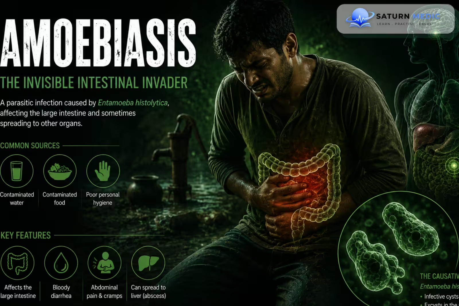
Amoebiasis is an intestinal infection caused by the protozoan Entamoeba histolytica. It is acquired mainly by ingesting contaminated food or water. Infection may be asymptomatic or cause diarrhea, dysentery, or an amoebic liver abscess
Also called
ICD-10
Specialty
Onset
Reviewed
-
OverviewOverview
-
Etiology & Risk FactorsEtiology & Risk Factors
-
PathophysiologyPathophysiology
-
Clinical PresentationClinical Presentation
-
History TakingHistory Taking
-
Physical ExaminationPhysical Examination
-
InvestigationsInvestigations
-
DiagnosisDiagnosis
-
ManagementManagement
-
ComplicationsComplications
-
PrognosisPrognosis
-
Key Points / Clinical PearlsKey Points / Clinical Pearls
Overview
-Amoebiasis may be asymptomatic or present as amoebic colitis or liver abscess.
–Intestinaldiseasecan cause abdominal pain, diarrhea, dysentery with blood and mucus, tenesmus, fever, and weight loss.
–Extraintestinalinfection most commonly affects the liver and may cause right upper quadrant pain, fever, tender hepatomegaly, and referred shoulder pain.
–Severe complications include fulminantcolitis,toxicmegacolon, intestinal perforation, peritonitis, and rupture of a liver abscess.
Etiology & Risk Factors
-Etiology
–Infection follows ingestion of mature E. histolytica cysts through contaminated food or water. Cysts survive the stomach, release trophozoites in the colon, and may invade the intestinal mucosa.
-Risk Factors
- Poor sanitation
- Contaminated food or water
- Travel or residence in endemic regions
- Oral-anal sexual exposure
- Immunosuppression
Pathophysiology
Ingestion of cysts → excystation → trophozoites colonize the colon → mucosal invasion → flask-shaped ulcers → dysentery ± portal spread → hepatic invasion → amoebic liver abscess
Clinical Presentation
-Symptoms:
-
Amoebiasis may cause:
- Abdominal cramps
- Diarrhea
- Bloody or mucoid stools
- Tenesmus
- Fatigue
- Weight loss
Amoebic Liver Abscess
- Fever
- Right upper quadrant pain
- Weight loss
- Hepatomegaly
-Signs:
-
Abdominal tenderness
-
Fever
-
Hepatomegaly in hepatic disease
-
Right upper quadrant tenderness
-
Severe Amoebiasis may cause fulminant colitis, toxic megacolon, intestinal perforation, severe bleeding, or ruptured liver abscess.
History Taking
-Ask about:
- Diarrhea duration and stool character
- Blood or mucus in stool
- Abdominal pain and tenesmus
- Fever and weight loss
- Travel or residence in endemic areas
- Unsafe food or water exposure
- Oral-anal sexual exposure
- Right upper quadrant pain
Physical Examination
-General Examination
- Temperature
- Hydration status
- Weight and nutritional status
-System-Specific Examination:
- Abdominal tenderness
- Hepatomegaly
- Right upper quadrant tenderness
- Peritonitis in severe disease
Investigations
-Complete Blood Count
Not routinely required in uncomplicated intestinal disease.
May show anemia or leukocytosis in severe disease.
Biochemistry / Specific Tests
-
Stool antigen testing or PCR when available
-
Stool microscopy for cysts and trophozoites
-
Liver function tests when hepatic disease is suspected
-
Serology for extraintestinal disease
Microscopy may not distinguish E. histolytica from nonpathogenic Entamoeba species.
-Imaging
Abdominal Ultrasound
Useful for detecting an amoebic liver abscess.
Computed Tomography (CT)
Used for complicated or unclear hepatic lesions.
-Special / Confirmatory Tests
For suspected liver abscess, E. histolytica serology can support the diagnosis.
Important Investigation Note
Stool microscopy alone may be insufficient because pathogenic E. histolytica can resemble nonpathogenic species.
Diagnosis
-Amoebiasis is diagnosed by compatible clinical findings plus detection of E. histolytica or supportive testing.
Dysentery → stool antigen/PCR or microscopy → identify E. histolytica → assess for extraintestinal disease
-For liver abscess:
Right upper quadrant symptoms + liver abscess on imaging → supportive serology
Related Topics
Management
Amoebiasis · Management
| Clinical situation | Treatment |
|---|---|
| Asymptomatic intestinal infection | Paromomycin, iodoquinol, or diloxanide furoate — luminal agent |
| Invasive intestinal disease Amoebic colitis | Metronidazole or tinidazole followed by a luminal agent |
| Amoebic liver abscess | Metronidazole or tinidazole followed by a luminal agent |
| Severe / complicated disease | Metronidazole or tinidazole + luminal agent; drainage may be required if abscess is large, ruptured, or not responding |
Complications
- Fulminant colitis
- Intestinal bleeding
- Toxic megacolon
- Intestinal perforation
- Peritonitis
- Amoebic liver abscess
- Abscess rupture
- Pleuropulmonary infection
- Brain abscess
- Death
Prognosis
The prognosis of Amoebiasis is generally good with appropriate treatment. Severe colitis and extraintestinal disease can be life-threatening, particularly when diagnosis is delayed or a liver abscess ruptures.
Key Points / Clinical Pearls
- Amoebiasis is caused by Entamoeba histolytica.
- Transmission occurs mainly by the fecal-oral route.
- Mature cysts are the infective form.
- Infection may be asymptomatic.
- Amoebic colitis can cause bloody diarrhea and tenesmus.
- The liver is the most common extraintestinal site.
- Liver abscess commonly causes fever and right upper quadrant pain.
- Stool microscopy may not distinguish pathogenic from nonpathogenic species.
- Stool antigen testing or PCR is more specific when available.
- Ultrasound is useful for suspected liver abscess.
- Metronidazole or tinidazole treats invasive disease.
- A luminal agent is required afterward.
- Complicated liver abscesses may require drainage.
- Centers for Disease Control and Prevention (CDC). Amebiasis .
- Shirley D-A T, Farr L, Watanabe K, Moonah S. A Review of the Global Burden, New Diagnostics, and Current Therapeutics for Amebiasis. Open Forum Infect Dis. 2018;5(7):ofy161. PubMed .
- Haque R, Huston CD, Hughes M, Houpt E, Petri WA Jr. Amebiasis. N Engl J Med. 2003;348(16):1565-1573. PubMed .
- Salles JM. Diagnosis and Treatment of Amebiasis: 100 Years of the State of the Art. Rev Soc Bras Med Trop. 2014;47(5):491-500. PubMed .