

Clinical Subject Page
Hydatid Cyst
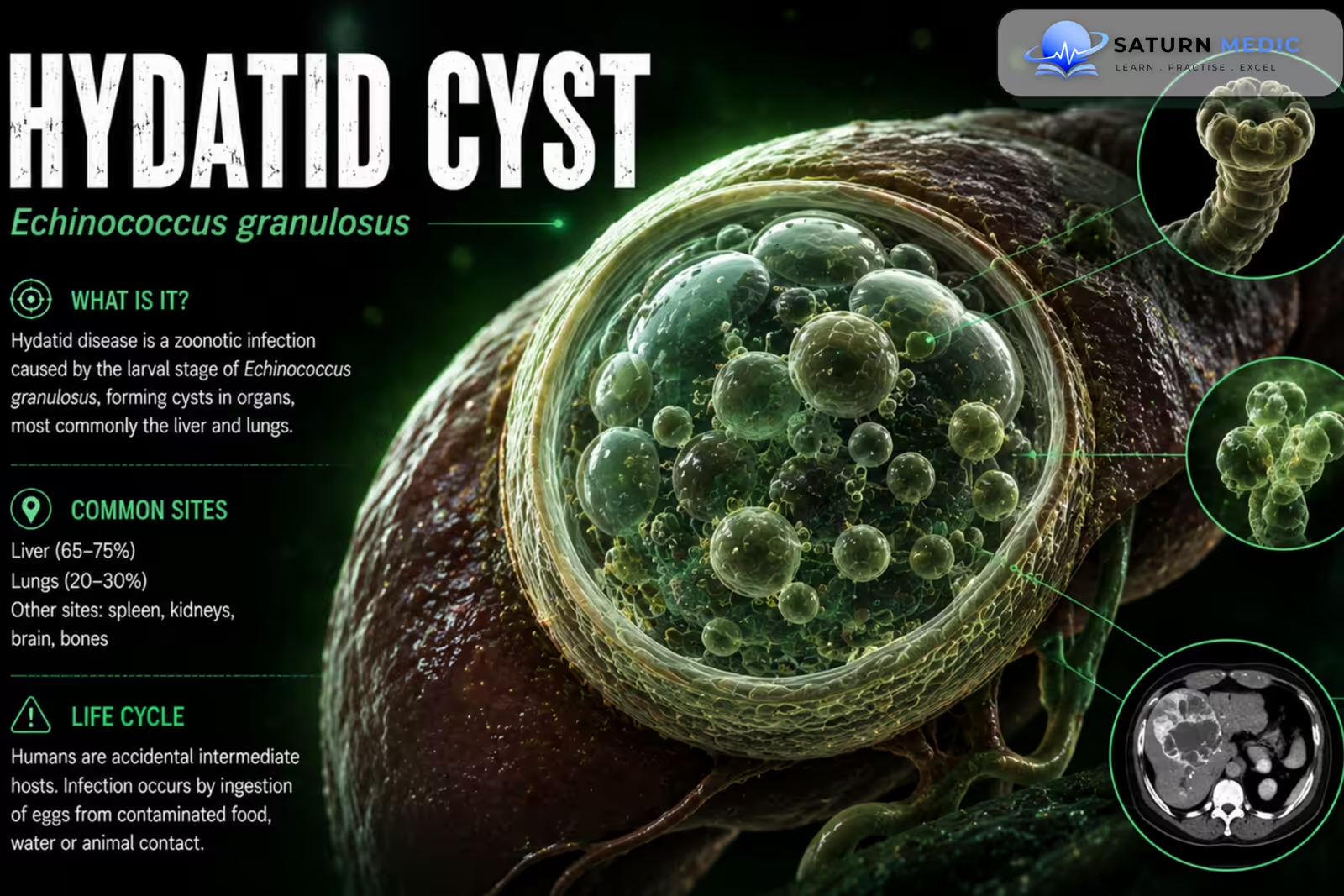
Hydatid Cyst is a parasitic cystic disease caused by infection with the larval stage of Echinococcus species, most commonly Echinococcus granulosus. Humans become accidental intermediate hosts after ingesting parasite eggs,The liver is the most commonly affected organ, followed by the lungs
Also called
ICD-10
Specialty
Onset
Reviewed
-
OverviewOverview
-
Etiology & Risk FactorsEtiology & Risk Factors
-
PathophysiologyPathophysiology
-
Clinical PresentationClinical Presentation
-
Life CycleLife Cycle
-
History TakingHistory Taking
-
Physical ExaminationPhysical Examination
-
InvestigationsInvestigations
-
DiagnosisDiagnosis
-
ManagementManagement
-
ComplicationsComplications
-
PrognosisPrognosis
-
Key Points / Clinical PearlsKey Points / Clinical Pearls
Overview
–Hydatid Cyst is a form of cystic echinococcosis. The parasite’s life cycle commonly involves dogs as definitive hosts and sheep or other livestock as intermediate hosts.
Humans acquire infection by ingesting eggs from contaminated food, water, or contact with infected dogs.
The main sites are:
- Liver
- Lungs
- Less commonly, kidneys, spleen, brain, bone, or other organs
Etiology & Risk Factors
-Etiology
–Hydatid Cyst is caused by the larval form of Echinococcus granulosus. After ingestion of eggs, embryos penetrate the intestinal wall and enter the portal circulation.
Most are filtered by the liver, while some reach the lungs or systemic circulation.
-Risk Factors
- Living in an endemic region
- Close contact with dogs
- Exposure to sheep or livestock
- Consumption of contaminated food or water
- Poor slaughterhouse or animal-handling practices
Pathophysiology
Ingestion of Echinococcus eggs → intestinal penetration → portal/systemic dissemination → larval development in an organ → slow-growing hydatid cyst → formation of daughter cysts and hydatid sand → enlargement → pressure effects or cyst complications
Clinical Presentation
-Symptoms:
A Hydatid Cyst may remain asymptomatic for years.
Symptoms depend on its location and size.
Hepatic Disease
- Right upper quadrant discomfort
- Abdominal fullness
- Hepatomegaly
- Nausea
- Obstructive jaundice if the biliary system is involved
Pulmonary Disease
- Cough
- Chest pain
- Dyspnea
- Hemoptysis
-Signs:
- Hepatomegaly
- Abdominal mass in large hepatic cysts
- Respiratory findings in pulmonary disease
- Jaundice when biliary obstruction occurs
Life Cycle
Hydatid Cyst · Echinococcus granulosus — Life Cycle
History Taking
-Ask about:
- Residence or travel in endemic areas
- Contact with dogs
- Contact with sheep or livestock
- Abdominal pain or fullness
- Cough or chest pain
- Dyspnea
- Hemoptysis
- Jaundice
- Previous hydatid disease
- Previous Hydatid Cyst surgery or treatment
Physical Examination
-General Examination
- Assess general condition
- Check for fever
- Look for allergic manifestations if rupture is suspected
-System-Specific Examination:
- Abdominal examination for hepatomegaly or mass
- Assess for jaundice
- Respiratory examination for pulmonary involvement
- Examine for signs of complications
Investigations
-Biochemistry / Specific Tests
Liver function tests when hepatic disease is suspected
Echinococcus-specific serology
Eosinophil count may support suspicion but cannot exclude disease when normal
-Imaging
Ultrasound
The preferred initial investigation for suspected hepatic Hydatid Cyst.
It may demonstrate:
Daughter cysts
Detached membranes
Hydatid sand
Calcification
Computed Tomography (CT)
Useful for defining cyst anatomy, calcification, complications, and disease extent.
Magnetic Resonance Imaging (MRI)
Useful for selected complex lesions and biliary or anatomical assessment.
-Special / Confirmatory Tests
Serology can support the diagnosis but has variable sensitivity depending on cyst location and stage.
Percutaneous aspiration should not be performed routinely for diagnosis because leakage can cause allergic reactions and dissemination.
Diagnosis
Hydatid Cyst is diagnosed by:
Exposure history + compatible cystic lesion → characteristic ultrasound/CT findings → supportive serology when appropriate → assessment for cyst complications and involvement of other organs.
The diagnosis is based on the combination of epidemiology, imaging, and laboratory findings.
Related Topics
Management
Hydatid Cyst · Surgical Priority — Case-Based Scenarios
| Rule | Principle | Rationale |
|---|---|---|
| 1st | Complicated cyst always first — ruptured, infected, or causing airway compromise | Life-threatening — anaphylaxis risk, sepsis, airway obstruction take priority over all else |
| 2nd | Lung before liver — when both are present and uncomplicated | Lung cysts rupture more easily, cause haemoptysis, bronchospasm, and anaphylaxis. Also — lung surgery first while liver compensates. Liver can wait longer safely. |
| 3rd | Right lung before left lung — when bilateral pulmonary cysts | Right lung is larger (3 lobes vs 2). Larger cyst volume = higher rupture risk. Also — right-sided thoracotomy easier; liver proximity makes right lung cysts more dangerous if they rupture into diaphragm. |
| 4th | Larger cyst before smaller — same organ, same side | Larger cysts: more tension, higher rupture risk, more daughter cysts |
| 5th | Right liver before left liver — bilateral hepatic cysts | Right lobe cysts more common and larger; closer to IVC and biliary tree — higher complication risk |
- Sterilise first — hypertonic saline 20% or H₂O₂ into cyst before opening
- Aspirate contents → open → remove endocyst
- Capitonnage — suture residual cavity walls together to obliterate dead space
- Bronchial fistulas closed individually
- Preserve lung tissue — avoid lobectomy unless destroyed
- PAIR (Puncture–Aspirate–Inject–Reaspitate) — minimally invasive; CE1/CE3a only
- Pericystectomy — removes pericyst + cyst; no residual cavity
- Omentoplasty — fill residual cavity with omentum
- Avoid PAIR if cyst communicates with biliary tree
- Always give albendazole 4 wks before + 4 wks after PAIR
- Protect field with hypertonic saline-soaked packs — kills protoscolices if spillage
- Never rupture cyst uncontrolled — anaphylaxis + seeding
- Sterilise before incising — inject hypertonic saline, wait 10–15 min
- Adrenaline + antihistamines + steroids ready — anaphylaxis kit
- Albendazole 400 mg BD — start 4 wks pre-op, continue 4 wks post-op
Complications
- Cyst rupture
- Anaphylaxis
- Secondary bacterial infection
- Biliary obstruction
- Obstructive jaundice
- Daughter-cyst dissemination
- Recurrent disease
- Pulmonary complications
Prognosis
The prognosis of Hydatid Cyst is generally good when the disease is diagnosed and treated appropriately. Outcome depends on cyst location, size, stage, complications, and completeness of treatment. Recurrence can occur, particularly after cyst spillage or incomplete treatment, so long-term imaging follow-up is important.
Key Points / Clinical Pearls
- Hydatid Cyst is caused by larval Echinococcus infection.
- Echinococcus granulosus is the most common cause of cystic echinococcosis.
- Dogs are important definitive hosts.
- Sheep and livestock commonly participate in the parasite’s life cycle.
- Humans acquire infection by ingesting parasite eggs.
- The liver is the most commonly affected organ.
- The lungs are the second most common site.
- Hydatid disease can remain asymptomatic for years.
- Daughter cysts are an important imaging feature.
- Ultrasound is particularly useful for hepatic disease.
- CT helps define cyst anatomy and complications.
- Serology supports but does not always confirm the diagnosis
- World Health Organization (WHO). Echinococcosis .
- Centers for Disease Control and Prevention (CDC). Echinococcosis .
- Brunetti E, Kern P, Vuitton DA; Writing Panel for the WHO-IWGE. Expert Consensus for the Diagnosis and Treatment of Cystic and Alveolar Echinococcosis in Humans. Acta Trop. 2010;114(1):1-16. PubMed .
- McManus DP, Gray DJ, Zhang W, Yang Y. Diagnosis, Treatment, and Management of Echinococcosis. BMJ. 2012;344:e3866. BMJ .
- Moro P, Schantz PM. Echinococcosis: A Review. Int J Infect Dis. 2009;13(2):125-133. PubMed .
- National Library of Medicine (NIH). Echinococcosis . StatPearls.