

Clinical Subject Page
Leishmaniasis
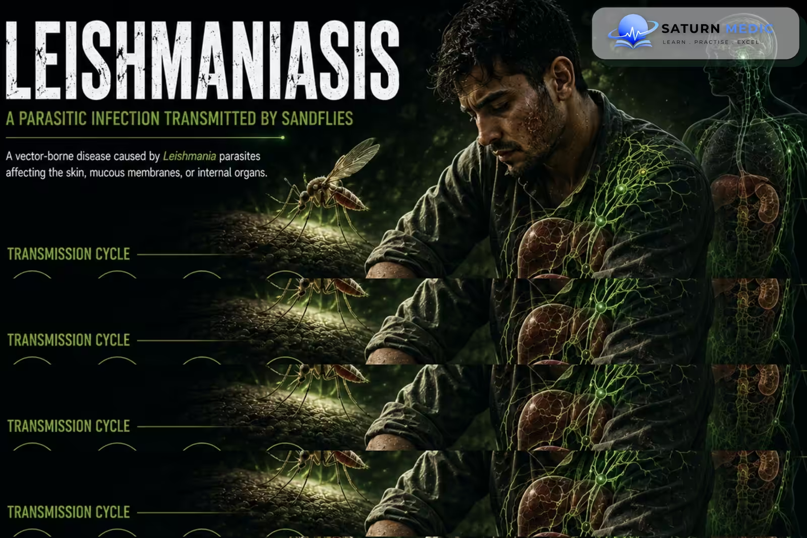
Leishmaniasis is a parasitic disease caused by Leishmania species and transmitted mainly through the bite of infected female sandflies. It ranges from localized skin disease to destructive mucosal disease and potentially fatal visceral infection
Also called
ICD-10
Specialty
Onset
Reviewed
-
OverviewOverview
-
Etiology & Risk FactorsEtiology & Risk Factors
-
PathophysiologyPathophysiology
-
Clinical PresentationClinical Presentation
-
Causative AgentsCausative Agents
-
History TakingHistory Taking
-
Physical ExaminationPhysical Examination
-
InvestigationsInvestigations
-
DiagnosisDiagnosis
-
ManagementManagement
-
ComplicationsComplications
-
PrognosisPrognosis
-
Key Points / Clinical PearlsKey Points / Clinical Pearls
Overview
–Leishmaniasis has three major clinical forms:
- Cutaneous: skin papules, nodules, or ulcers
- Mucocutaneous: destructive lesions of the nasal and oral mucosa
- Visceral: systemic infection involving the spleen, liver, and bone marrow
Visceral disease is the most severe form and may be fatal if untreated
Etiology & Risk Factors
-Etiology
–Leishmaniasis is caused by intracellular protozoa of the genus Leishmania.
The parasite exists as:
- Amastigotes: intracellular form found within macrophages
- Promastigotes: form transmitted by the sandfly
Transmission occurs mainly through infected female phlebotomine sandflies.
-Risk Factors
- Residence or travel in endemic areas
- Sandfly exposure
- Immunosuppression
- HIV infection
- Malnutrition
Pathophysiology
Sandfly bite → promastigotes enter tissue → uptake by macrophages → transformation into amastigotes → intracellular multiplication → local tissue damage or systemic dissemination → cutaneous, mucocutaneous, or visceral disease
Clinical Presentation
-Symptoms:
Clinical features of Leishmaniasis depend on the form.
Cutaneous
- Papule or nodule at the bite site
- Gradual enlargement
- Painless ulcer
- Raised ulcer margins
Mucocutaneous
- Nasal obstruction
- Epistaxis
- Nasal or oral ulceration
- Progressive mucosal destruction
Visceral
- Prolonged fever
- Weight loss
- Weakness
- Loss of appetite
- Abdominal fullness
-Signs:
- Chronic skin ulcer or nodule
- Mucosal ulceration
- Splenomegaly
- Hepatomegaly
- Pallor
- Lymphadenopathy in some patients
Severe visceral disease may cause:
- Pancytopenia
- Severe anemia
- Bleeding
- Secondary bacterial infection
- Severe wasting
- Hypotension
- Organ dysfunction
Causative Agents
Leishmania · Causative Agents
| Species | Disease | Classic clue | Key association |
|---|---|---|---|
| L. donovani | Visceral | Kala-azar | Fever + massive splenomegaly + pancytopenia |
| L. infantum | Visceral | Zoonotic visceral disease | Dogs = important reservoir |
| L. tropica | Cutaneous | Dry lesion | Old World · urban |
| L. major | Cutaneous | Wet ulcer | Old World · rodents |
| L. braziliensis | Mucocutaneous | Mucosal destruction | New World · South/Central America |
History Taking
-Ask about:
- Residence or travel in endemic areas
- Sandfly exposure
- Duration and progression of skin lesions
- Fever and weight loss
- Nasal or oral symptoms
- Abdominal fullness
- Previous Leishmaniasis
- Immunosuppression or HIV infection
Physical Examination
-General Examination
- Temperature
- Weight and nutritional status
- Pallor
- Signs of chronic illness
-System-Specific Examination:
- Inspect skin lesions
- Examine nasal and oral mucosa
- Palpate the spleen and liver
- Assess lymph nodes
- Look for bleeding or secondary infection
Investigations
-Complete Blood Count
Important in visceral disease and may show:
-
Anemia
-
Leukopenia
-
Thrombocytopenia
-
Pancytopenia
-Biochemistry / Specific Tests
Depending on the clinical form:
-
Liver function tests
-
Renal function tests
-
Leishmania serology, particularly for visceral disease
-
Polymerase Chain Reaction (PCR) where available
-
HIV testing when clinically appropriate
-Imaging
-
Ultrasound may demonstrate hepatosplenomegaly in visceral disease.
-
Further imaging is guided by organ involvement or suspected complications.
-Special / Confirmatory Tests
Tissue or Bone Marrow Examination
Demonstration of Leishmania amastigotes in tissue can confirm visceral disease.
For cutaneous disease, microscopy, culture, or PCR from lesion material may establish the diagnosis.
Diagnosis
-Diagnosis of Leishmaniasis combines epidemiological exposure, clinical findings, and detection of the parasite or its DNA.
Endemic exposure + compatible clinical syndrome → targeted serology or tissue/lesion testing → identify Leishmania → determine clinical form and severity
Related Topics
Management
1. First-Line / Emergency Management
There is no routine emergency treatment for uncomplicated cutaneous disease.
Severe visceral disease requires urgent specialist treatment, particularly with:
-
Severe anemia
-
Bleeding
-
Secondary infection
-
Hemodynamic instability
2. Definitive Treatment
Treatment depends on:
-
Clinical form
-
Leishmania species
-
Geographic region
-
Disease severity
-
Immune status
Cutaneous disease may require local or systemic treatment, while mucocutaneous and visceral disease generally require systemic therapy.
3. Medical Treatment
Important treatments include:
-
Liposomal amphotericin B — important treatment for visceral disease
-
Miltefosine — used for selected species and clinical forms
-
Pentavalent antimonials in selected settings
-
Local therapy for selected cutaneous lesions
4. Supportive Management
-
Nutritional support
-
Treatment of anemia and cytopenias
-
Treatment of secondary infection
-
HIV assessment and treatment when relevant
Complications
- Secondary bacterial infection
- Permanent skin scarring
- Mucosal destruction
- Epistaxis
- Pancytopenia
- Severe anemia
- Bleeding
- Relapse
- Post-kala-azar dermal disease
- Death in untreated visceral disease
Prognosis
The prognosis of Leishmaniasis depends on the clinical form, species, immune status, and treatment. Cutaneous disease may heal but can leave permanent scars. Mucocutaneous disease may cause progressive tissue destruction. Visceral disease is potentially fatal without treatment but can usually be cured with appropriate therapy
Key Points / Clinical Pearls
- Leishmaniasis is caused by Leishmania protozoa.
- It is transmitted mainly by infected female sandflies.
- The three major forms are cutaneous, mucocutaneous, and visceral.
- Visceral disease is also called kala-azar.
- Cutaneous disease commonly causes a chronic painless ulcer.
- Mucocutaneous disease can destroy nasal and oral tissues.
- Visceral disease commonly causes prolonged fever, weight loss, and splenomegaly.
- Pancytopenia is an important feature of visceral disease.
- Diagnosis depends on the clinical form.
- Serology is particularly useful in visceral disease.
- Detection of amastigotes or parasite DNA can confirm infection.
- Liposomal amphotericin B is an important treatment for visceral disease.
- Miltefosine is used for selected species and clinical forms.
- World Health Organization (WHO). Leishmaniasis .
- Centers for Disease Control and Prevention (CDC). Leishmaniasis .
- Aronson N, Herwaldt BL, Libman M, et al. Diagnosis and Treatment of Leishmaniasis: Clinical Practice Guidelines by the Infectious Diseases Society of America (IDSA) and the American Society of Tropical Medicine and Hygiene (ASTMH). Clin Infect Dis. 2016;63(12):e202-e264. PubMed .
- Burza S, Croft SL, Boelaert M. Leishmaniasis. Lancet. 2018;392(10151):951-970. PubMed .