

Clinical Subject Page
Rabies
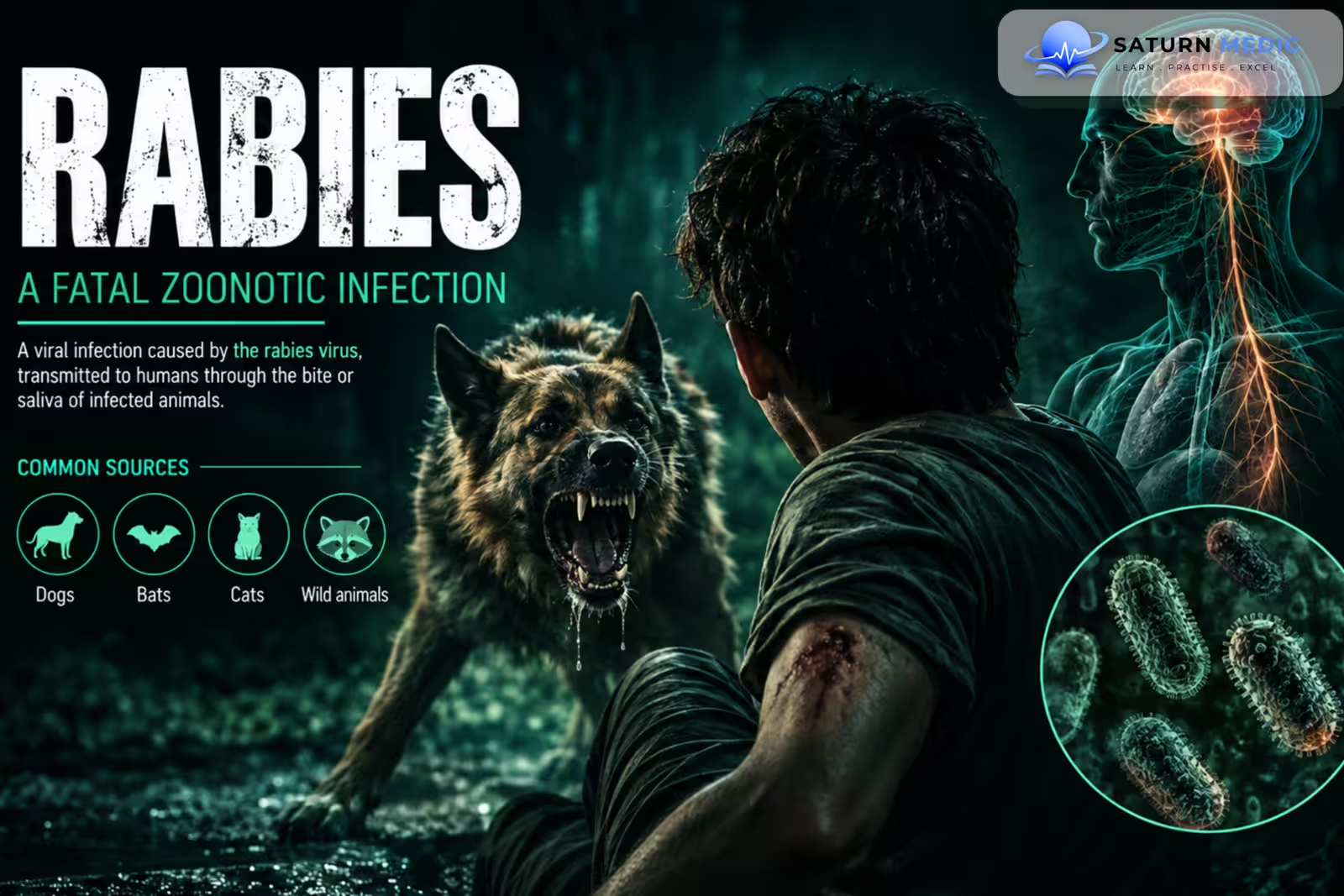
Rabies is a fatal viral infection of the central nervous system caused by the rabies virus. It is a zoonotic disease transmitted mainly through the saliva of infected mammals, most commonly through bites. Once clinical neurological symptoms develop, the disease is almost invariably fatal, but prompt post-exposure prophylaxis can prevent disease before symptoms begin
Also called
ICD-10
Specialty
Onset
Reviewed
-
OverviewOverview
-
Etiology & Risk FactorsEtiology & Risk Factors
-
PathophysiologyPathophysiology
-
Clinical PresentationClinical Presentation
-
History TakingHistory Taking
-
Physical ExaminationPhysical Examination
-
InvestigationsInvestigations
-
DiagnosisDiagnosis
-
ManagementManagement
-
ComplicationsComplications
-
PrognosisPrognosis
-
Key Points / Clinical PearlsKey Points / Clinical Pearls
Overview
–Rabies primarily affects the brain and spinal cord after the virus travels from the site of inoculation through peripheral nerves toward the central nervous system.
Human infection is usually caused by bites or scratches from infected mammals. Dogs are responsible for the vast majority of human cases globally.
-The disease has two major clinical patterns:
- Furious rabies: agitation, hydrophobia, hallucinations, and autonomic instability.
- Paralytic rabies: progressive flaccid paralysis resembling Guillain-Barré syndrome.
Etiology & Risk Factors
-Etiology
–Rabies is caused by rabies virus, an enveloped, single-stranded RNA virus of the Rhabdoviridae family.
The virus is introduced into tissue through infected saliva and initially replicates locally before entering peripheral nerves.
-Risk Factors
- Bite or scratch from a potentially infected mammal
- Exposure to unvaccinated dogs in endemic areas
- Contact with bats
- Occupational animal exposure
- Living in or traveling to rabies-endemic regions
Pathophysiology
Animal bite or scratch → viral inoculation into tissue → local viral replication → entry into peripheral nerves → retrograde axonal transport → central nervous system infection → encephalitis → autonomic and neurological dysfunction → coma and death
Clinical Presentation
-Symptoms:
Early symptoms include:
- Fever
- Headache
- Weakness
- Malaise
- Pain, tingling, or itching at the bite site
Later symptoms may include:
- Anxiety
- Agitation
- Confusion
- Hallucinations
- Difficulty swallowing
- Hydrophobia
- Aerophobia
- Excessive salivation
-Signs:
- Fever
- Agitation or altered mental status
- Pharyngeal spasms
- Hypersalivation
- Autonomic instability
- Progressive neurological dysfunction
- Encephalitis
- Generalized spasms
- Paralysis
- Respiratory failure
History Taking
-Ask about:
- Animal bite or scratch
- Species and behavior of the animal
- Date and location of exposure
- Whether saliva contacted broken skin or mucosa
- Whether the animal was available for observation or testing
- Previous rabies vaccination
- Previous post-exposure prophylaxis
- Travel or residence in an endemic area
- Onset of fever or neurological symptoms
Physical Examination
-General Examination
- Temperature
- Mental status
- Hydration
- Cardiovascular and respiratory status
- Signs of autonomic dysfunction
-System-Specific Examination:
- Neurological examination
- Assessment for pharyngeal spasms
- Assessment for cranial nerve abnormalities
- Assessment for progressive weakness or paralysis
- Examination of the exposure wound
Investigations
-Biochemistry / Specific Tests
Testing is mainly required when clinical disease is suspected.
Serum and cerebrospinal fluid testing for rabies-specific antibodies
Molecular detection of viral RNA by Polymerase Chain Reaction (PCR) from appropriate specimens
Routine biochemical tests to assess organ dysfunction in severe disease
-Imaging
Not routinely required to diagnose Rabies.
Brain imaging may be used to exclude alternative neurological diagnoses.
-Special / Confirmatory Tests
Diagnosis of clinical Rabies requires specialized laboratory testing coordinated with public-health authorities. Testing may involve saliva, nuchal skin biopsy, cerebrospinal fluid, or serum depending on the clinical stage.
Important Investigation Note
Post-exposure prophylaxis should never be delayed while waiting for diagnostic testing when a significant exposure has occurred.
Diagnosis
-Clinical Rabies is suspected in a patient with:
Compatible animal exposure → prodromal symptoms → progressive encephalitis, hydrophobia, or paralysis.
-Laboratory diagnosis is performed using specialized PCR and antibody testing, but management of an exposure is based primarily on the exposure risk rather than waiting for laboratory confirmation.
Related Topics
Management
Rabies · Post-Exposure Prophylaxis (PEP)
| Step | Action | Critical rule |
|---|---|---|
| Step 1 Wound wash |
Immediate and thorough washing with soap and water for 15 minutes minimum — most important single intervention. Then irrigate with povidone-iodine or 70% alcohol. | Do this BEFORE going to hospital. Reduces viral load at entry site. Never suture immediately — delay or avoid primary closure. |
| Step 2 Risk assess |
Category I: touching/feeding animal, licks on intact skin → no PEP needed. Category II: nibbling, minor scratches without bleeding → vaccine only. Category III: transdermal bites, scratches with bleeding, licks on broken skin, mucous membrane exposure → vaccine + RIG. |
WHO exposure categories I / II / III — determines PEP regimen |
| Step 3 Rabies immunoglobulin (RIG) |
Human RIG (HRIG) 20 IU/kg — infiltrate as much as possible around wound; remainder IM at distant site. Equine RIG (ERIG) 40 IU/kg — if HRIG unavailable; skin test first. Give on day 0 only — never after day 7. |
Category III only. Give with 1st vaccine dose — different site. Provides immediate passive immunity while vaccine response develops. |
| Step 4 Rabies vaccine |
Previously unvaccinated: 4 doses — days 0, 3, 7, 14 (IM deltoid) Previously vaccinated: 2 doses only — days 0 + 3 (no RIG needed) Vaccine: cell-culture vaccine (HDCV, PCECV) — not nerve tissue vaccines |
Never inject in gluteal — poor immune response. Deltoid only (thigh in infants). Start as soon as possible — no upper time limit if exposure confirmed. |
Rabies · Management of Established Disease & Supportive Care
| Problem | Management | Notes |
|---|---|---|
| Agitation / spasms | Midazolam infusion — 1st line sedation Diazepam IV PRN for breakthrough spasms Ketamine — anaesthetic doses for refractory agitation |
Dark, quiet room — minimise stimuli triggering spasms (same as tetanus) |
| Hydrophobia / aerophobia | Deep sedation during episodes. NG tube — if oral route impossible. IV fluids for hydration. | Never force oral fluids — precipitates fatal laryngospasm |
| Respiratory failure | Mechanical ventilation — intubation early if paralytic form or declining GCS. Tracheostomy for prolonged ventilation. | May prolong survival but does not alter outcome once symptomatic |
| Autonomic instability | Cardiac monitoring — arrhythmias common. Labetalol — sympathetic surges. Atropine — bradycardia/vagal episodes. |
Leading cause of death in ICU-managed patients |
| Pain / comfort | Morphine infusion — analgesia + reduces sympathetic surges. Palliative intent in most cases. | Goal shifts to comfort when prognosis confirmed |
| Milwaukee protocol | Induced coma (ketamine/midazolam) + antiviral agents (ribavirin/amantadine) + supportive ICU care. Used in the only well-documented survivor (Jeanna Giese, 2004). | Largely abandoned — replicated in <5 survivors out of hundreds of attempts. Not standard of care. |
Complications
- Encephalitis
- Hydrophobia
- Respiratory failure
- Autonomic instability
- Cardiac arrhythmias
- Paralysis
- Coma
- Multiorgan dysfunction
- Death
Prognosis
The prognosis of Rabies depends critically on whether post-exposure prophylaxis is given before symptoms develop. Properly administered prophylaxis is highly effective at preventing disease. Once clinical neurological symptoms appear, Rabies is almost invariably fatal, and treatment is primarily supportive.
Key Points / Clinical Pearls
- Rabies is a fatal viral infection of the central nervous system.
- It is caused by rabies virus.
- Transmission occurs mainly through the saliva of infected mammals.
- Dog bites account for most human cases globally.
- The virus travels through peripheral nerves to the central nervous system.
- Incubation is usually weeks to months.
- Early symptoms are nonspecific and may include fever and headache.
- Hydrophobia and painful pharyngeal spasms are characteristic features.
- A paralytic form can present with progressive weakness and paralysis.
- There is no reliable curative treatment once clinical disease develops.
- Immediate wound washing for at least 15 minutes is essential after a potentially infectious exposure.
- Post-exposure prophylaxis includes vaccine and, when indicated, rabies immunoglobulin.
- Previously vaccinated patients require a different PEP regimen.
- World Health Organization (WHO). Rabies .
- Centers for Disease Control and Prevention (CDC). Rabies .
- World Health Organization (WHO). WHO Expert Consultation on Rabies: Third Report . Technical Report Series 1012.
- Hemachudha T, Laothamatas J, Rupprecht CE. Human Rabies: A Disease of Complex Neuropathogenetic Mechanisms and Diagnostic Challenges. Lancet Neurol. 2002;1(2):101-109. PubMed .
- Fooks AR, Cliquet F, Finke S, et al. Rabies. Nat Rev Dis Primers. 2017;3:17091. PubMed .
- Manning SE, Rupprecht CE, Fishbein D, et al. Human Rabies Prevention — United States, 2008: Recommendations of the Advisory Committee on Immunization Practices. MMWR Recomm Rep. 2008;57(RR-3):1-28. CDC .
- National Library of Medicine (NIH). Rabies . StatPearls.