

Clinical Subject Page
Staphylococcal Infection
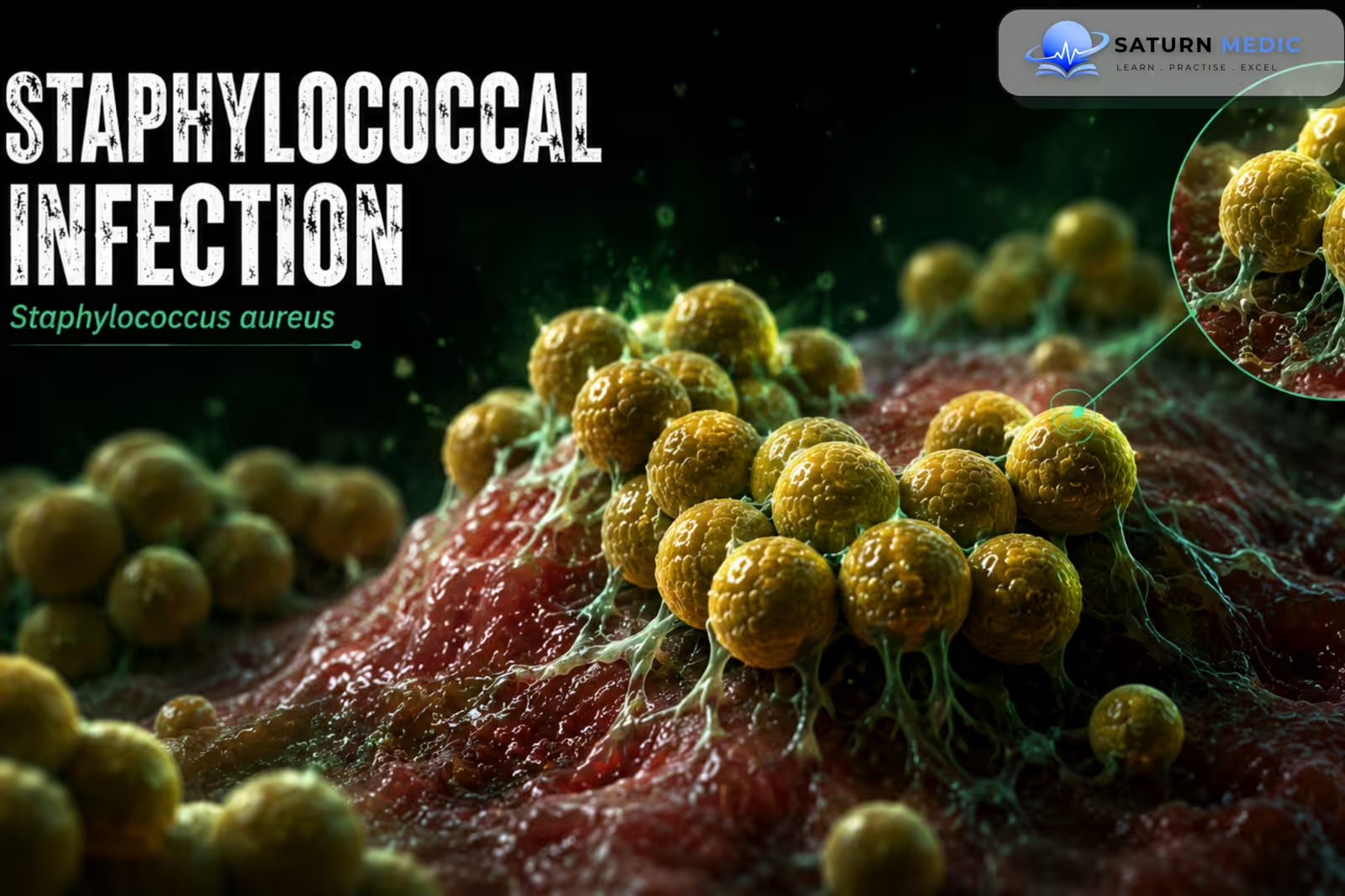
Staphylococcal Infection is an infection caused by bacteria of the genus Staphylococcus. The most clinically important species is Staphylococcus aureus, which can cause infections ranging from localized skin disease to life-threatening bacteremia, endocarditis,pneumonia, osteomyelitis, and sepsis.
Also called
ICD-10
Specialty
Onset
Reviewed
-
OverviewOverview
-
Etiology & Risk FactorsEtiology & Risk Factors
-
PathophysiologyPathophysiology
-
Clinical PresentationClinical Presentation
-
Bacterial ClassificationBacterial Classification
-
History TakingHistory Taking
-
Physical ExaminationPhysical Examination
-
InvestigationsInvestigations
-
DiagnosisDiagnosis
-
ManagementManagement
-
ComplicationsComplications
-
PrognosisPrognosis
-
Key Points / Clinical PearlsKey Points / Clinical Pearls
Overview
–Staphylococcal Infection can affect almost any body system.
–S. aureus commonly colonizes the skin and anterior nares without causing disease, but it can enter damaged tissue or the bloodstream and produce invasive infection.
–Common clinical manifestations include:
- Skin and soft-tissue infections
- Abscesses
- Wound infections
- Bacteremia
- Endocarditis
- Osteomyelitis
- Septic arthritis
- Pneumonia
- Toxic shock syndrome
- Food poisoning
Etiology & Risk Factors
-Etiology
–Staphylococcal Infection is most commonly caused by Staphylococcus aureus, a Gram-positive, catalase-positive, coagulase-positive coccus that commonly forms clusters.
Transmission occurs through:
- Direct skin-to-skin contact
- Contact with contaminated surfaces
- Autoinoculation from colonized skin or mucosa
-Risk Factors
- Skin breaks or wounds
- Recent surgery or hospitalization
- Indwelling catheters or medical devices
- Diabetes mellitus
- Immunosuppression
Pathophysiology
Colonization → disruption of skin or mucosal barrier → bacterial invasion → local inflammation and tissue destruction → abscess or invasive infection → bloodstream dissemination → sepsis and metastatic infection
Clinical Presentation
-Symptoms:
Presentation depends on the site of infection.
Skin and Soft Tissue
- Painful skin lesion
- Redness
- Swelling
- Warmth
- Pus or drainage
- Fever
Invasive Infection
- Fever
- Chills
- Malaise
- Hypotension
- Altered mental status
- Shortness of breath
Endocarditis
- Persistent fever
- Fatigue
- New or changing heart murmur
- Embolic manifestations
-Signs:
- Erythematous, warm, tender skin lesion
- Fluctuant abscess
- Purulent drainage
- Fever
- Tachycardia
- Hypotension in severe infection
Bacterial Classification
Staphylococci · Classification
| Factor | Key mechanism | Exam association |
|---|---|---|
| Protein A | Binds Fc of IgG → blocks opsonisation | Immune evasion |
| Coagulase | Fibrin formation around bacteria | Coagulase-positive Staph |
| Exfoliative toxin | Cleaves desmoglein-1 | SSSS + bullous impetigo |
| TSST-1 | Superantigen → cytokine release | Toxic Shock Syndrome |
| Enterotoxin | Preformed, heat-stable toxin | Food poisoning — rapid vomiting |
| PVL | Leukocyte lysis | Necrotising skin infection / pneumonia |
History Taking
-Ask about:
- Onset and progression of symptoms
- Skin wounds or abscesses
- Recent surgery
- Recent hospitalization
- Intravenous catheter or other medical devices
- Previous MRSA infection
- Antibiotic exposure
- Diabetes or immunosuppression
- Intravenous drug use
- Joint or bone pain
Physical Examination
-General Examination
- Temperature
- Heart rate and blood pressure
- General appearance
- Signs of sepsis or shock
-System-Specific Examination:
- Inspect skin for abscesses and wounds
- Examine joints for swelling and restricted movement
- Examine bones for localized tenderness
- Cardiac examination for murmurs
- Respiratory examination for pneumonia
- Examine catheter and surgical sites
Investigations
-Complete Blood Count
Useful for assessing systemic infection and inflammatory response.
-Biochemistry / Specific Tests
-
Blood cultures for suspected bacteremia or systemic infection
-
Culture and antimicrobial susceptibility testing from purulent material
-
Serum creatinine and other organ-function tests in systemic infection
-
Inflammatory markers when useful for monitoring
-Imaging
Imaging depends on the suspected site:
-
Ultrasound for suspected abscess
-
Echocardiography when endocarditis is suspected
-
X-ray or Magnetic Resonance Imaging (MRI) for suspected bone or joint infection
-
Chest imaging for suspected pneumonia
-Special / Confirmatory Tests
Bacterial Culture
Culture confirms the organism and allows antimicrobial susceptibility testing.
Testing is particularly important for distinguishing:
-
MSSA (Methicillin-susceptible Staphylococcus aureus)
- MRSA (Methicillin-resistant Staphylococcus aureus)
Important Investigation Note
Blood cultures are essential in suspected invasive Staphylococcus aureus infection, while purulent collections should be cultured whenever possible.
Diagnosis
Staphylococcal Infection is diagnosed by:
Compatible clinical syndrome → appropriate specimen collection → bacterial culture → identification of Staphylococcus species → antimicrobial susceptibility testing.
For suspected bacteremia, blood cultures are essential. The infection source should then be identified and assessed for metastatic spread.
Related Topics
Management
Staphylococcal Infections · Treatment by Condition
| Condition | 1st Line | Alternative | Duration |
|---|---|---|---|
| Skin & soft tissue Cellulitis, furunculosis, abscess |
Flucloxacillin 500 mg QDS PO Abscess: incision & drainage — antibiotics only if surrounding cellulitis |
Co-amoxiclav PO Cefalexin 500 mg QDS PO |
5–7 days |
| Impetigo Bullous (S. aureus) or non-bullous |
Topical mupirocin — mild/localised Flucloxacillin PO — extensive |
Cefalexin PO | 5–7 days |
| Pneumonia Community / post-influenza |
Flucloxacillin 1–2g IV q6h | Cefazolin 2g IV q8h Clindamycin if penicillin allergy |
7–14 days |
| Bacteraemia / Endocarditis | Flucloxacillin 2g IV q4h — gold standard for MSSA endocarditis | Cefazolin 2g IV q8h — non-inferior, less CNS penetration | 4–6 weeks (endocarditis) 14 days (bacteraemia) |
| Osteomyelitis / Septic arthritis | Flucloxacillin 2g IV q6h → step-down to PO when improving | Cefalexin or Clindamycin PO (step-down) | 4–6 weeks (osteo) 3–4 weeks (septic arthritis) |
| Toxic Shock Syndrome (TSS) | Flucloxacillin 2g IV q4h + Clindamycin 900 mg IV q8h — clindamycin suppresses toxin production | IV immunoglobulin (IVIG) — adjunct for refractory TSS | 10–14 days |
| Scalded Skin Syndrome (SSSS) | Flucloxacillin IV → PO Supportive: fluid/electrolyte replacement, wound care |
Cefalexin PO (mild) | 7–10 days |
| Food poisoning Preformed enterotoxin |
Supportive only — oral rehydration. Antibiotics NOT indicated (preformed toxin — no live bacteria to treat) | Self-limiting 24–48h | |
| Condition | 1st Line | Alternative | Duration |
|---|---|---|---|
| Skin & soft tissue (mild) CA-MRSA, abscess, cellulitis |
Co-trimoxazole (TMP-SMX) PO — CA-MRSA 1st line Doxycycline 100 mg BD PO |
Clindamycin PO — check D-zone test for inducible resistance Abscess: I&D alone often sufficient |
5–7 days |
| Skin & soft tissue (severe) Necrotising fasciitis, severe cellulitis |
Vancomycin IV — target trough 15–20 mg/L or AUC/MIC 400–600 + Clindamycin — anti-toxin effect (PVL) |
Linezolid 600 mg BD IV/PO | 7–14 days |
| Pneumonia Necrotising / cavitating |
Linezolid 600 mg BD IV — preferred over vancomycin (better lung penetration) + Clindamycin — suppresses PVL toxin |
Vancomycin IV — if linezolid not available | 7–21 days |
| Bacteraemia | Vancomycin IV — gold standard Echocardiogram to exclude endocarditis |
Daptomycin 6–10 mg/kg IV OD — non-inferior; good for persistent bacteraemia | 14 days minimum (longer if endocarditis) |
| Endocarditis | Vancomycin IV — 6 weeks native valve; 6 weeks + rifampicin + gentamicin for prosthetic | Daptomycin 8–10 mg/kg IV — left-sided endocarditis alternative | 6 weeks |
| Osteomyelitis / Septic arthritis | Vancomycin IV → step-down to PO agent when sensitive Step-down: TMP-SMX or Linezolid PO |
Daptomycin IV — alternative to vancomycin + Rifampicin — adjunct for biofilm penetration |
4–6 weeks (osteo) |
| Meningitis / CNS | Linezolid IV — best CNS penetration Vancomycin IV — standard; adjust dose for CSF levels |
TMP-SMX IV — adequate CNS penetration | 14–21 days |
| Decolonisation Pre-op / carriers |
Mupirocin 2% nasal ointment BD × 5 days Chlorhexidine 4% body wash daily × 5 days |
Octenidine — if mupirocin resistance Add oral antibiotics only if systemic carriage |
5 days |
| Condition | Treatment | Notes |
|---|---|---|
| Prosthetic valve endocarditis | Vancomycin IV + Rifampicin + Gentamicin (first 2 weeks) | Often requires valve replacement surgery |
| Central line / catheter infection | Vancomycin IV — remove line if possible Line salvage: vancomycin lock therapy |
Remove device whenever feasible — biofilm = antibiotic failure |
| Joint prosthesis infection | Vancomycin IV + Rifampicin Step-down: Rifampicin + TMP-SMX or doxycycline PO |
Debridement + implant retention (DAIR) or two-stage revision |
| CSF shunt infection | Vancomycin IV ± intrathecal vancomycin Shunt removal + external drainage |
Shunt removal essential for cure — antibiotics alone fail |
Complications
- Abscess formation
- Bacteremia
- Sepsis
- Septic shock
- Endocarditis
- Pneumonia
- Osteomyelitis
- Septic arthritis
- Metastatic abscesses
- Toxic shock syndrome
- Multiorgan failure
Prognosis
The prognosis varies widely according to the site and severity of infection. Localized skin infections generally have a good outcome with appropriate treatment and source control. Bacteremia, endocarditis, pneumonia, and other invasive infections carry substantially greater morbidity and mortality, particularly when treatment or source control is delayed.
Key Points / Clinical Pearls
- Staphylococcal Infection is most commonly caused by Staphylococcus aureus.
- S. aureus commonly colonizes the skin and nose.
- It can cause both localized and invasive disease.
- Skin and soft-tissue infections are common manifestations.
- Abscesses commonly require incision and drainage.
- MSSA and MRSA require different antibiotic considerations.
- Culture and susceptibility testing guide definitive antibiotic therapy.
- Blood cultures are essential when bacteremia is suspected.
- S. aureus bacteremia requires evaluation for a deep infection source.
- Endocarditis is an important complication of bacteremia.
- Osteomyelitis and septic arthritis can occur through hematogenous spread.
- Infected medical devices may require removal.
- Source control is a key component of treatment.
- Centers for Disease Control and Prevention (CDC). Staphylococcus aureus and Staph Infections .
- Tong SYC, Davis JS, Eichenberger E, Holland TL, Fowler VG Jr. Staphylococcus aureus Infections: Epidemiology, Pathophysiology, Clinical Manifestations, and Management. Clin Microbiol Rev. 2015;28(3):603-661. PubMed .
- Lowy FD. Staphylococcus aureus Infections. N Engl J Med. 1998;339(8):520-532. New England Journal of Medicine .
- Murray CJ, et al. Global Burden of Bacterial Antimicrobial Resistance in 2019: A Systematic Analysis. Lancet. 2022;399(10325):629-655. PubMed .
- Stevens DL, Bisno AL, Chambers HF, et al. Practice Guidelines for the Diagnosis and Management of Skin and Soft Tissue Infections. Clin Infect Dis. 2014;59(2):e10-e52. Clinical Infectious Diseases .
- Liu C, Bayer A, Cosgrove SE, et al. Clinical Practice Guidelines by the Infectious Diseases Society of America for the Treatment of Methicillin-Resistant Staphylococcus aureus Infections in Adults and Children. Clin Infect Dis. 2011;52(3):e18-e55. PubMed .
- National Library of Medicine (NIH). Staphylococcus aureus Infection . StatPearls.