

Clinical Subject Page
Viral Hemorrhagic Fever (VHF)
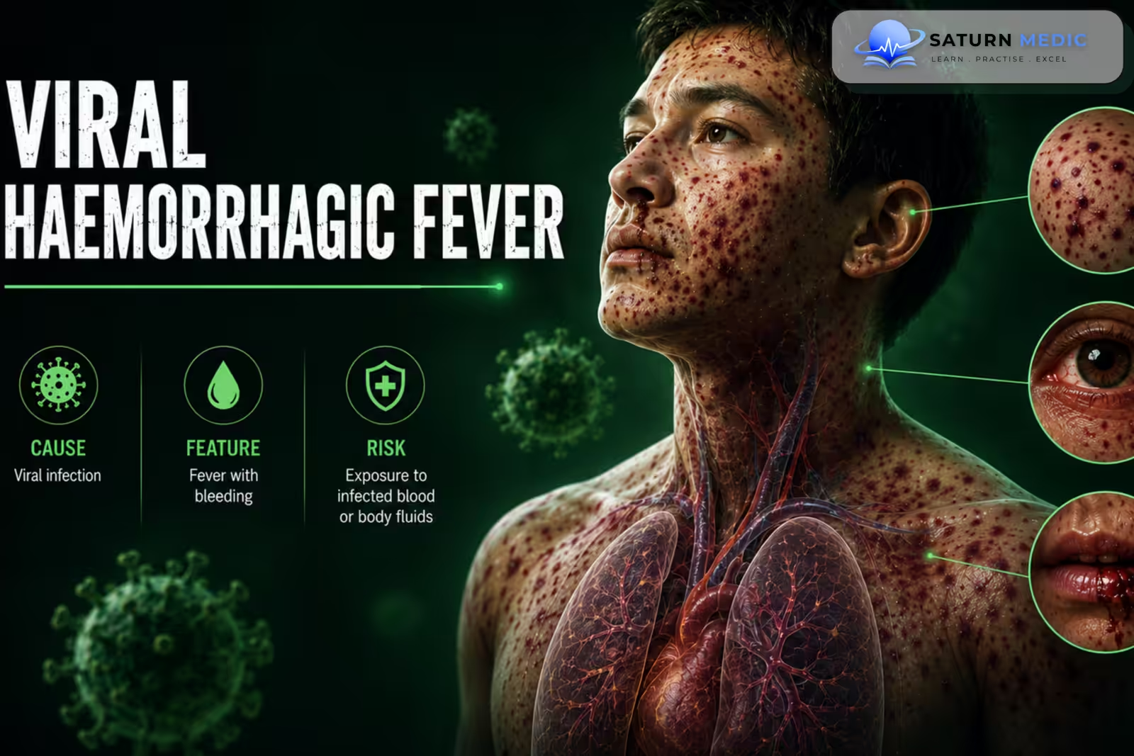
Viral Hemorrhagic Fever (VHF) is a group of severe viral illnesses that can cause fever, vascular dysfunction, bleeding, and multi-organ involvement. Important causes include Ebola, Marburg, Lassa, and Crimean-Congo hemorrhagic fever
Also called
ICD-10
A99
Specialty
Onset
Reviewed
-
OverviewOverview
-
Etiology & Risk FactorsEtiology & Risk Factors
-
PathophysiologyPathophysiology
-
Clinical PresentationClinical Presentation
-
Types of VHF VirusesTypes of VHF Viruses
-
History TakingHistory Taking
-
Physical ExaminationPhysical Examination
-
InvestigationsInvestigations
-
DiagnosisDiagnosis
-
ManagementManagement
-
ComplicationsComplications
-
PrognosisPrognosis
-
Key Points / Clinical PearlsKey Points / Clinical Pearls
Overview
–Viral Hemorrhagic Fever (VHF) is a syndrome caused by viruses from several families.
-Important examples include:
- Ebola and Marburg viruses
- Lassa virus
- Crimean-Congo hemorrhagic fever virus
- Some arenaviruses
- Dengue and yellow fever viruses
-Transmission varies and may occur through mosquitoes or ticks, infected animals, contaminated food or materials, or direct contact with infected body fluids
Etiology & Risk Factors
-Etiology
–The viruses causing Viral Hemorrhagic Fever (VHF) have different reservoirs and transmission routes:
- Mosquito or tick exposure
- Contact with infected rodents, bats, or livestock
- Exposure to infected blood or body fluids
- Contact with infected patients or contaminated materials
-Risk Factors
- Residence or travel in an endemic area
- Contact with infected animals
- Healthcare exposure
- Laboratory exposure
- Close contact with infected patients
Pathophysiology
Viral infection → systemic viral replication → endothelial and immune dysfunction → increased vascular permeability + coagulation abnormalities → tissue hypoperfusion and organ injury → bleeding in some patients → shock and multi-organ failure
Clinical Presentation
-Symptoms:
Early Viral Hemorrhagic Fever (VHF) is often nonspecific:
- Fever
- Severe headache
- Myalgia
- Weakness and fatigue
- Nausea and vomiting
- Diarrhea
- Abdominal pain
-Signs:
- Fever
- Rash in some infections
- Dehydration
- Hypotension
- Tachycardia
- Petechiae or mucosal bleeding
-Severe Disease
- Hemorrhagic manifestations
- Shock
- Acute Kidney Injury (AKI)
- Liver dysfunction
- Encephalopathy
- Disseminated Intravascular Coagulation (DIC)
- Respiratory failure
- Multi-organ failure
Types of VHF Viruses
Viral Haemorrhagic Fevers · Types by Virus Family
| Virus | Species / Strains | Reservoir | Transmission | Mortality | Endemic region |
|---|---|---|---|---|---|
| Ebola virus | 5 species: Zaire (most lethal), Sudan, Bundibugyo, Taï Forest, Reston (non-pathogenic in humans) | Fruit bats (Pteropodidae) — probable. Spillover via bushmeat. | Direct contact — blood, body fluids, secretions. No airborne transmission. | 25–90% (Zaire highest) |
Sub-Saharan Africa (DRC, Guinea, Sierra Leone, Uganda) |
| Marburg virus | Single species — Marburg marburgvirus. Two strains: Marburg + Ravn. | Egyptian fruit bat (Rousettus aegyptiacus) — confirmed reservoir | Direct contact with blood/fluids. Bat caves (miners, tourists). Healthcare nosocomial. | 24–88% | Central/East Africa (Uganda, DRC, Angola, Guinea). Linked to bat caves. |
| Virus | Transmission | Reservoir | Clinical hallmark | Mortality | Region |
|---|---|---|---|---|---|
| CCHF Crimean-Congo HF |
Hyalomma tick bite — primary. Livestock blood/tissue. Nosocomial (healthcare workers). | Hyalomma ticks + livestock (cattle, sheep, goats) — amplifying hosts | 3 phases: pre-haemorrhagic → haemorrhagic (petechiae, ecchymoses, GI bleed) → convalescence. Hepatitis common. | 5–40% | Africa, Middle East (Iraq), Balkans, Central/South Asia |
| Hantavirus HFRS / HPS |
Rodent aerosol — inhalation of infected urine/faeces/saliva. No person-to-person (except Andes virus). | Rodents — species-specific: Hantaan (Apodemus), Seoul (Rattus), Sin Nombre (deer mouse) | HFRS (Old World): fever + renal failure + haemorrhage. HPS (New World): severe pulmonary oedema — "hantavirus pulmonary syndrome" | HFRS: 1–15% HPS: 30–40% |
HFRS: Asia, Europe. HPS: Americas |
| Rift Valley Fever | Mosquito bite (Aedes, Culex). Contact with infected animal blood/abortion material. Aerosol from slaughterhouses. | Livestock (sheep, cattle, goats) — amplifying hosts. Mosquitoes — vector. | Most mild febrile illness. Severe (<2%): retinitis, encephalitis, haemorrhagic fever. High mortality in neonatal animals (abortion storms). | <1% overall Up to 50% severe |
Sub-Saharan Africa, Egypt, Arabian Peninsula |
| SFTS Severe Fever with Thrombocytopenia |
Haemaphysalis tick bite. Limited person-to-person via blood/fluids. | Ticks (Haemaphysalis longicornis). Hedgehogs, wild animals — suspected reservoir. | Fever + thrombocytopenia + leukopenia + GI symptoms. CNS involvement in severe cases. | 12–30% | China, South Korea, Japan |
| Virus | Reservoir | Transmission | Clinical hallmark | Mortality | Region |
|---|---|---|---|---|---|
| Lassa virus | Mastomys natalensis (multimammate rat) | Rodent urine/faeces contaminating food. Person-to-person via blood/fluids. Nosocomial. | 80% asymptomatic/mild. Severe: facial oedema, pleural effusion, haemorrhage, deafness (30% survivors). Ribavirin effective. | 1–15% overall 50% hospitalised |
West Africa (Nigeria, Sierra Leone, Guinea, Liberia) |
| Junin virus Argentine HF |
Calomys musculinus (corn mouse) | Rodent aerosol/contact during harvest. Occupational — farm workers. | Haemorrhage + neurological signs (tremor, seizures). Seasonal — harvest time. | 15–30% (untreated) |
Argentina (pampas region) |
| Machupo virus Bolivian HF |
Calomys callosus (vesper mouse) | Rodent contact. Limited person-to-person. | Similar to Junin — haemorrhage + neurological features. | 25–35% | Bolivia |
| Lujo virus | Unknown rodent reservoir | Person-to-person nosocomial (high efficiency — index case → 4 secondary cases) | Severe VHF with rash. Fatal in 4/5 of initial outbreak. | 80% | Southern Africa (Zambia, South Africa) |
| Sabia virus Brazilian HF |
Unknown reservoir | Unknown natural transmission. Lab accidents documented. | Severe haemorrhagic fever. Very few cases described. | High | Brazil |
| Virus | Vector | Reservoir | Clinical hallmark | Mortality | Region |
|---|---|---|---|---|---|
| Yellow Fever | Aedes aegypti (urban), Haemagogus (sylvatic) | Primates (jungle cycle). Humans — urban cycle amplifying host. | Remission phase (deceptive recovery) → Intoxication phase (jaundice + haemorrhage + Faget's sign). Black vomit (haematemesis). | Mild: <5% Severe: 20–50% |
Sub-Saharan Africa, South America. Preventable by 17D vaccine. |
| Dengue virus Severe dengue |
Aedes aegypti, Aedes albopictus | Humans are the primary amplifying host (urban). Primates (sylvatic cycle). | 4 serotypes. Secondary infection (different serotype) → antibody-dependent enhancement → severe dengue: plasma leak, haemorrhage, shock (dengue shock syndrome). Tourniquet test positive. | <1% (treated) Up to 20% untreated |
Tropical/subtropical worldwide. Most common arboviral disease. |
| Kyasanur Forest Disease (KFD) | Haemaphysalis tick | Hard ticks + small mammals (rodents, monkeys) | Biphasic illness: fever + haemorrhage → remission → neurological phase (meningitis, tremors). Monkey deaths = sentinel event. | 3–5% | India (Karnataka state). Vaccine available. |
| Omsk HF | Dermacentor tick. Contact with muskrats. | Muskrats, water voles, ticks | Biphasic: fever + haemorrhage → neurological features (encephalitis). Milder than KFD. | 0.5–3% | Western Siberia (Russia) |
| Alkhurma HF | Tick bite (Ornithodoros). Sheep/camel slaughter. | Camels, sheep — amplifying hosts. Ticks — vector. | Fever + haemorrhage + encephalitis. Related to KFD. | ~25% | Saudi Arabia |
History Taking
-Ask about:
- Fever onset and progression
- Bleeding or bruising
- Headache and myalgia
- Vomiting or diarrhea
- Travel or residence in endemic regions
- Mosquito or tick exposure
- Contact with rodents, bats, livestock, or primates
- Contact with suspected or confirmed cases
- Healthcare or laboratory exposure
Physical Examination
-General Examination
- Temperature
- Blood pressure
- Heart rate
- Respiratory rate and oxygen saturation
- Mental status
- Hydration and perfusion
-System-Specific Examination:
- Inspect skin and mucosa for bleeding
- Assess the abdomen and neurological status
- Look for shock and organ dysfunction
Investigations
-Complete Blood Count
Useful for assessing:
Thrombocytopenia
Leukopenia or leukocytosis
Anemia
-Biochemistry / Specific Tests
Serum electrolytes
Urea and creatinine
Liver function tests
Coagulation profile
Serum lactate
Blood cultures when bacterial sepsis is a differential
Virus-specific PCR or serology when appropriate
-Imaging
Imaging is guided by symptoms and suspected complications.
Chest imaging for respiratory involvement
Ultrasound or CT for suspected bleeding or organ complications
-Special / Confirmatory Tests
Reverse Transcription Polymerase Chain Reaction (RT-PCR) is an important diagnostic method for many VHFs. Antigen detection and serology may also be used depending on the suspected virus.
Laboratory specimens require strict biosafety and infection-control procedures.
Diagnosis
- Hemorrhage
- Shock
- Acute Kidney Injury (AKI)
- Disseminated Intravascular Coagulation (DIC)
- Encephalitis
- Respiratory failure
- Multi-organ failure
- Death
Related Topics
Management
Viral Haemorrhagic Fevers · Overview & Key VHFs
up to 50% hospitalised
Viral Haemorrhagic Fevers · Supportive Care & Infection Control
| Problem | Management | Notes |
|---|---|---|
| Fluid loss / shock | IV crystalloid (normal saline / Ringer's lactate) — aggressive replacement matching losses. Oral rehydration if tolerated. Central line if massive losses — use ultrasound guidance, minimise trauma. | Lassa: cautious — pulmonary oedema risk. Ebola: 5–10L/day losses common. |
| Electrolyte imbalance | Monitor K⁺, Na⁺, Mg²⁺, Ca²⁺ — correct aggressively. Hypokalaemia + hypomagnesaemia common from diarrhoea. | Electrolyte correction improves outcomes significantly |
| Haemorrhage / DIC | Fresh frozen plasma (FFP) — replenish clotting factors. Platelet transfusion if <50,000 + active bleeding. Cryoprecipitate for fibrinogen <1g/L. Tranexamic acid — consider for severe mucosal bleeding. | Avoid aspirin, NSAIDs, heparin. Packed RBC if Hb <7 + haemodynamically compromised. |
| Fever / pain | Paracetamol — antipyretic + analgesia. Avoid NSAIDs / aspirin — inhibit platelet function, worsen bleeding. | Morphine PO/IV for severe pain if needed |
| Nausea / vomiting | Ondansetron IV/PO. Metoclopramide — caution if CNS involved. Maintain NG feeding if oral route lost. | Anti-emetics critical — GI losses are primary driver of shock |
| Secondary infections | Empirical broad-spectrum antibiotics if bacterial sepsis suspected. Treat malaria — always co-test in endemic areas (malaria + VHF can co-exist). | Malaria rapid test on all febrile patients from endemic regions |
| Renal failure | Monitor urine output. Avoid nephrotoxic drugs. Dialysis — if AKI refractory. Fluid balance strict monitoring. | AKI common in Yellow fever, Hantavirus, severe Ebola |
| Encephalopathy | Correct metabolic causes (hypoglycaemia, electrolytes). Seizure management: benzodiazepines. Avoid LP if coagulopathic — bleeding risk. | Common in Lassa, severe Ebola |
Complications
- Encephalitis
- Meningitis
- Pneumonitis
- Hepatitis
- Retinitis
- Neonatal infection
- Disseminated infection
- Malignancy
Prognosis
The prognosis varies with the virus, disease severity, comorbidities, and access to early supportive care. Some VHFs are self-limited, whereas Ebola, Marburg, and severe Crimean-Congo hemorrhagic fever can be highly fatal. Early recognition and supportive care improve outcomes
Key Points / Clinical Pearls
- Viral Hemorrhagic Fever (VHF) is a syndrome caused by several different viruses.
- Important causes include Ebola, Marburg, Lassa, and Crimean-Congo hemorrhagic fever.
- Transmission differs between viruses.
- Exposure history is essential in suspected cases.
- Early symptoms are usually nonspecific.
- Fever, headache, myalgia, vomiting, and diarrhea are common.
- Bleeding may occur but is not always present.
- Thrombocytopenia and coagulation abnormalities may occur in severe disease.
- RT-PCR is important for many VHFs.
- Suspected cases require immediate infection-control precautions.
- Public-health notification is essential.
- Treatment is mainly supportive for many VHFs.
- Some VHFs have disease-specific therapies or vaccines.
- World Health Organization (WHO). Viral Haemorrhagic Fevers .
- Centers for Disease Control and Prevention (CDC). Viral Hemorrhagic Fevers .
- Centers for Disease Control and Prevention (CDC). About Viral Hemorrhagic Fevers .
- Kortepeter MG, Bausch DG, Bray M. Basic Clinical and Laboratory Features of Filoviral Hemorrhagic Fevers. J Infect Dis. 2011;204 Suppl 3:S810-S816. PubMed .
- Paessler S, Walker DH. Pathogenesis of the Viral Hemorrhagic Fevers. Annu Rev Pathol. 2013;8:411-440. PubMed .
- Fauci AS, Lane HC. Viral Hemorrhagic Fevers. In: Harrison's Principles of Internal Medicine.
- National Library of Medicine (NIH). Viral Hemorrhagic Fevers . StatPearls.