

Clinical Subject Page
Alpha-1 Antitrypsin Deficiency (AATD)
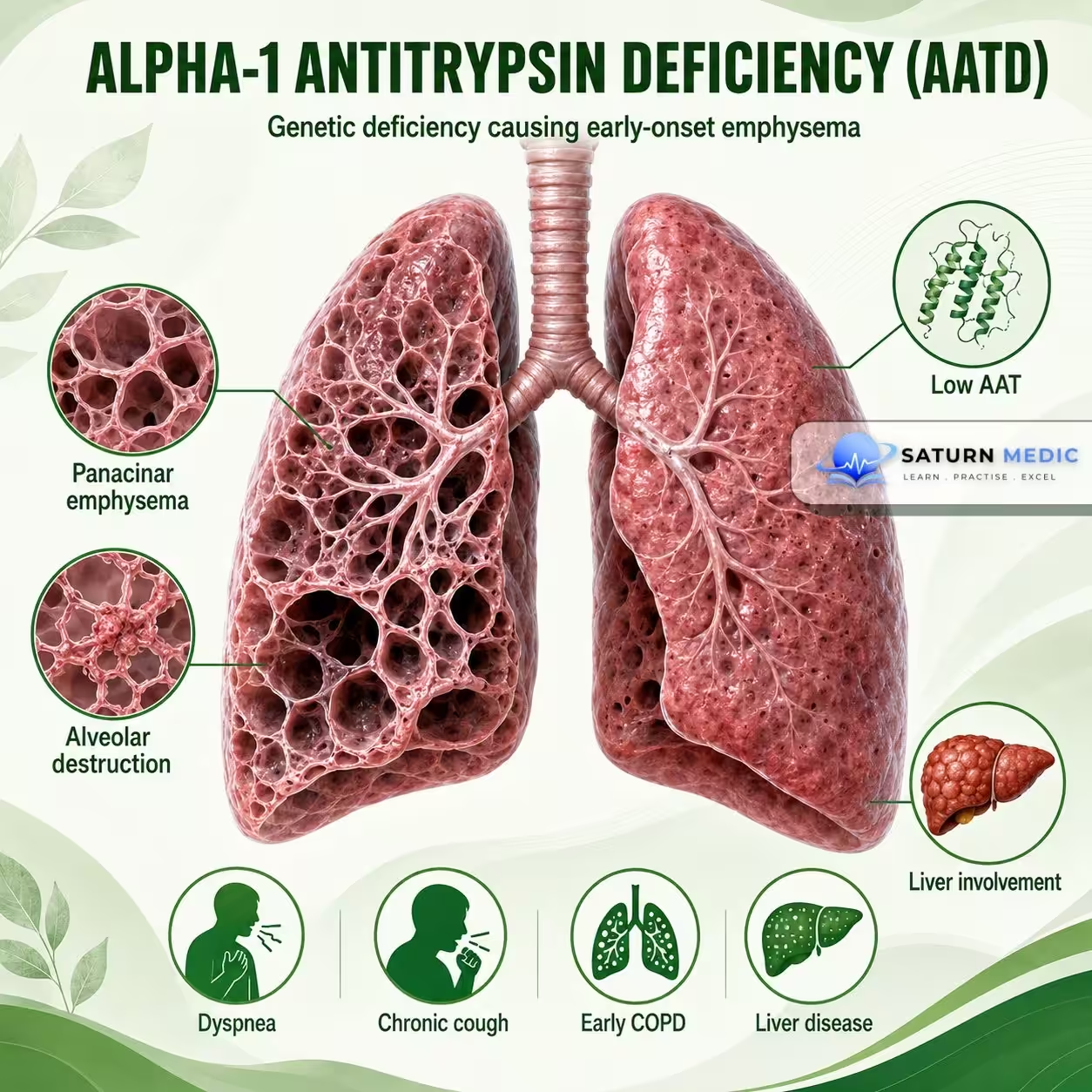
Alpha-1 Antitrypsin Deficiency :An inherited genetic disorder causing low levels of alpha-1 antitrypsin (AAT), a protein that protects lung tissue from neutrophil elastase. Its deficiency leads to early-onset emphysema (especially in the lower lobes) and may also cause liver disease.
Also called
Alpha-1 proteinase inhibitor deficiency
ICD-10
E88.01
Specialty
Pulmonology
Onset
Chronic
Reviewed
July 2026
On This Page
-
OverviewOverview
-
Etiology & Risk FactorsEtiology & Risk Factors
-
PathophysiologyPathophysiology
-
Clinical PresentationClinical Presentation
-
History TakingHistory Taking
-
Physical ExaminationPhysical Examination
-
InvestigationsInvestigations
-
DiagnosisDiagnosis
-
ManagementManagement
-
ComplicationsComplications
-
PrognosisPrognosis
-
Key Points / Clinical PearlsKey Points / Clinical Pearls
Overview
Alpha-1 Antitrypsin is a protein mainly produced by the liver. It protects the lungs from destructive enzymes, especially neutrophil elastase.
Deficiency causes:
- Lungs: Loss of protection → Alveolar destruction → Emphysema
- Liver: Abnormal AAT accumulation → Liver injury
Etiology & Risk Factors
-Causes of Alpha-1 Antitrypsin Deficiency
Alpha-1 Antitrypsin Deficiency is caused by mutations in the SERPINA1 gene.
-Inheritance
Autosomal codominant
Important Alleles
M allele: Normal
Z allele: Most important severe deficiency allele
S allele: Causes milder deficiency
Severe Disease
The PiZZ genotype is strongly associated with severe AAT deficiency.
Risk Factors for Lung Disease
Cigarette smoking
Occupational dust exposure
Air pollution
Recurrent respiratory infections
Important Note
Smoking greatly accelerates lung damage in patients with Alpha-1 Antitrypsin Deficiency
Pathophysiology
-Lung Disease
SERPINA1 mutation
⬇
↓ Functional alpha-1 antitrypsin
⬇
Unopposed neutrophil elastase activity
⬇
Destruction of alveolar walls
⬇
Loss of elastic recoil
⬇
Emphysema + Persistent airflow obstruction
-Liver Disease
Abnormal AAT protein production
⬇
Protein accumulation inside hepatocytes
⬇
Liver cell injury
⬇
Fibrosis
⬇
Cirrhosis and possible liver cancer
Key Concept
Lung disease results from protein deficiency, while liver disease results from abnormal protein accumulation.
Clinical Presentation
-Respiratory Features
- Progressive dyspnea
- Wheezing
- Chronic cough
- Sputum production
- Reduced exercise tolerance
- Recurrent respiratory infections
- Early-onset emphysema
-Typical Lung Pattern
Emphysema often:
- Develops at a younger age
- Is worse in smokers
- Predominantly affects the lower lungs
-Liver Features
- Neonatal jaundice
- Elevated liver enzymes
- Chronic hepatitis
- Cirrhosis
- Liver failure
-Other Features
- Panniculitis — rare
- Granulomatosis with polyangiitis association
History Taking
-Ask about:
- Shortness of breath?
- Chronic cough?
- Wheezing?
- Exercise limitation?
- Age when symptoms started?
- Smoking history?
- Occupational exposure?
- Previous diagnosis of COPD or emphysema?
- Liver disease or jaundice?
- Family history of emphysema?
- Family history of liver disease?
- Previous AAT testing?
Physical Examination
-Respiratory Examination
Look for:
Tachypnea
Prolonged expiration
Pursed-lip breathing
Hyperinflated chest
Reduced breath sounds
Wheezing
Advanced Lung Disease
Cyanosis
Accessory muscle use
Reduced oxygen saturation
-Liver Examination
Look for:
Jaundice
Hepatomegaly
Splenomegaly
Ascites
Signs of chronic liver disease
Investigations
-Serum Alpha-1 Antitrypsin Level — Initial Test
A low serum AAT level suggests the diagnosis.
-Genotyping or Phenotyping
Used to:
Confirm the diagnosis
Identify the specific AAT variant
Assess disease risk
-Pulmonary Function Tests
May show:
Obstructive airflow pattern
Reduced FEV₁/FVC ratio
Reduced gas transfer
-Chest CT
May show:
Emphysema
Predominantly lower-lung involvement
-Liver Assessment
Liver function tests
Liver ultrasound
Other fibrosis assessment when indicated
Diagnosis
-Diagnosis is based on:
1. Reduced Serum AAT Level
Together with:
2. Confirmation of the Abnormal Variant
Using:
- Genotyping
- Phenotyping
Important Note
AAT levels alone may be misleading because alpha-1 antitrypsin can increase during inflammation.
Related Topics
Management
Alpha-1 Antitrypsin Deficiency (AATD) · Management
| Treatment | Indication / Details | Notes |
|---|---|---|
| Smoking Cessation | All patients — absolute priority | Smokers with AATD lose lung function 5–10x faster than non-smokers. Varenicline, NRT, behavioural support. No other intervention is as effective. |
| AAT Augmentation Therapy | IV Prolastin / Respreeza (human pooled AAT) weekly | Indicated for PiZZ (or other severe genotypes) with established airflow obstruction (FEV1 35–65% predicted in most guidelines). Slows CT density loss (emphysema marker). Does not help liver disease. Lifelong weekly infusion. |
| Bronchodilators | LABA + LAMA (e.g. formoterol + tiotropium) | Same as COPD management — mainstay for symptomatic airflow obstruction. SABA as reliever. LABA/LAMA combination preferred for moderate-severe disease. |
| Inhaled Corticosteroids | ICS + LABA if frequent exacerbations or asthmatic overlap | Not recommended routinely in pure emphysematous AATD. Consider triple therapy (ICS/LABA/LAMA) only in exacerbation-prone patients or eosinophilic phenotype. |
| Pulmonary Rehabilitation | MRC dyspnoea grade ≥3 | Improves exercise capacity, quality of life, and reduces exacerbations. All patients with symptomatic lung disease should be referred. |
| Vaccinations | All patients | Annual influenza, pneumococcal (PCV13 + PPSV23), COVID-19, hepatitis A and B (especially if liver disease present). |
| Long-term Oxygen Therapy (LTOT) | PaO2 ≤7.3 kPa or SpO2 ≤88% at rest | Criteria same as COPD-related LTOT. ≥15 hours/day. Reduces pulmonary hypertension and improves survival in hypoxaemic patients. |
| Liver Transplantation | End-stage liver disease | Curative for the genetic defect — the donor liver produces normal AAT. Post-transplant serum AAT normalises. Does not reverse pre-existing lung damage but prevents further liver-mediated Z-protein accumulation. |
| Lung Transplantation | End-stage emphysema (FEV1 <25%) | Bilateral lung transplant preferred. Corrects lung disease but does not alter underlying genetic defect — recipient still produces Z-protein from native liver. Augmentation therapy stops post-transplant. |
| Liver Monitoring | Annual LFTs, ultrasound ± fibroscan | All PiZZ patients need annual liver surveillance. Ursodeoxycholic acid and alcohol avoidance. Refer to hepatology if fibrosis/cirrhosis detected. Screen for hepatocellular carcinoma if cirrhotic. |
| Family Screening & Genetic Counselling | All index cases | Autosomal codominant inheritance. Screen first-degree relatives with serum AAT level + Pi genotyping. Offer genetic counselling re: reproductive implications. Register with national AATD registry where available. |
Complications
- Early-onset emphysema
- COPD
- Respiratory failure
- Pulmonary hypertension
- Cor pulmonale
- Cirrhosis
- Liver failure
- Hepatocellular carcinoma
- Panniculitis
- Death
Prognosis
- Prognosis varies widely.
- The major factors affecting prognosis include:
- Smoking
- Severity of deficiency
- Degree of lung damage
- Presence of liver disease
- Smokers develop lung disease earlier and more rapidly.
- Early diagnosis and smoking avoidance can significantly improve outcomes.
- Advanced lung or liver disease may require transplantation.
Key Points / Clinical Pearls
- Alpha-1 Antitrypsin Deficiency is caused by mutations in the SERPINA1 gene.
- It mainly affects the lungs and liver.
- Lung disease results from unopposed neutrophil elastase activity.
- Liver disease results from abnormal AAT accumulation in hepatocytes.
- Consider it in young patients with early emphysema.
- Lower-lung predominant emphysema is a classic pattern.
- Smoking dramatically accelerates lung damage.
- Low serum AAT is followed by genotyping or phenotyping for confirmation.
- Meseeha M, Sankari A, Attia M. National Center for Biotechnology Information (NIH). Alpha-1 Antitrypsin Deficiency, StatPearls.
- Stoller JK, Kim V, Silverman EK, Sandhaus RA. National Center for Biotechnology Information (NIH). Alpha-1 Antitrypsin Deficiency, GeneReviews.
- Sandhaus RA, Turino G, Brantly ML, et al. The Diagnosis and Management of Alpha-1 Antitrypsin Deficiency in the Adult. Chronic Obstr Pulm Dis. 2016. PMID: 28848868.
- MedlinePlus, National Library of Medicine (NIH). Alpha-1 Antitrypsin Deficiency: Genetics Home Reference.
- National Center for Biotechnology Information (NIH). Emphysema, StatPearls.